Showing posts with label Physiology. Show all posts

March 30, 2023

HEART OR PULSE RATE


INTRODUCTION: A healthy adult has a resting heart rate or pulse of 60 to 80 beats per minute, which is the rate of depolarization of the SA node. It should be noted that, the SA node actually has a slightly faster rate, closer to 100 beats per minute, but is slowed by parasympathetic nerve impulses to what we consider a normal resting rate.

PARAMETERS OF HEART RATE: A rate less than 60, except for athletes, is called bradycardia; a prolonged or consistent rate greater than 100 beats per minute is called tachycardia. A child’s normal heart rate may be as high as 100 beats per minute, that of an infant as high as 120, and that of a near-term fetus as high as 140 beats per minute. These higher rates are not related to age, but rather to size: the smaller the individual, the higher the metabolic rate and the faster the heart rate.

INTERPRETATION OF PULSE RATE: A pulse is the heartbeat that is felt at an arterial site. What is felt is not actually the force exerted by the blood, but the force of ventricular contraction transmitted through the walls of the arteries. This is why pulses are not felt in veins; they are too far from the heart for the force to be detectable. The most commonly used pulse sites are:

Radial; the radial artery on the thumb side of the wrist.

Carotid; the carotid artery lateral to the larynx in the neck.

Temporal; the temporal artery just in front of the ear.

Femoral; the femoral artery at the top of the thigh.

Popliteal; the popliteal artery at the back of the knee.

Dorsalis pedis; the dorsalis pedis artery on the top of the foot, commonly called the pedal pulse.

Pulse rate is, of course, the heart rate. However, if the heart is beating weakly, a radial pulse may be lower than an apical pulse. This is called a pulse deficit and indicates heart disease of some kind.

CONCLUSION: When taking a pulse, the careful observer also notes the rhythm and force of the pulse. Abnormal rhythms may reflect cardiac arrhythmias, and the force of the pulse (strong or weak) is helpful in assessing the general condition of the heart and arteries.

RELATED;

1.  CARDIAC OUTPUT

2. HEART MURMURS

3.  HEART FAILURE

REERENCES

March 24, 2023

OVULATION AND THE MENSTRUAL CYCLE

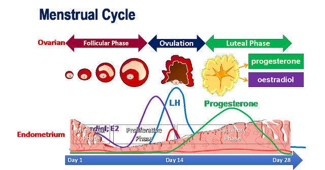
INTRODUCTION: The menstrual cycle includes the activity of the hormones of the ovaries and anterior pituitary gland and the resultant changes in the ovaries that we sometimes call, the ovarian cycle. This then is preceded by changes in the uterus which we sometimes call the uterine cycle.

HORMONES INVOLVED IN THE PROCESS: Follicle Stimulating (FSH) and Luteinizing Hormone (LH) from the anterior pituitary gland, estrogen from the ovarian follicle, and progesterone from the corpus luteum are the four hormones involved. It is the fluctuation and production of these hormones that influence the ovulation cycle and and then menstruations.

THE CYCLE: A cycle may be described in terms of three phases: menstrual phase, follicular phase, and luteal phase.

1. Menstrual phase: The loss of the functional layer of the endometrium is called menstruation or the menses. Although this is actually the end of a menstrual cycle, the onset of menstruation is easily pinpointed and is, therefore, a useful starting point. Menstruation may last 2 to 8 days, with an average of 3 to 6 days. At this time, secretion of FSH is increasing, and several ovarian follicles begin to develop.

2. Follicular phase: FSH stimulates growth of ovarian follicles and secretion of estrogen by the follicle cells. The secretion of LH is also increasing, but more slowly. FSH and estrogen promote the growth and maturation of the ovum, and estrogen stimulates the growth of blood vessels in the endometrium to regenerate the functional layer. This phase ends with ovulation, when a sharp increase in LH causes rupture of a mature ovarian follicle.

3. Luteal phase: Under the influence of LH, the ruptured follicle becomes the corpus luteum and begins to secrete progesterone as well as estrogen. Progesterone stimulates further growth of blood vessels in the functional layer of the endometrium and promotes the storage of nutrients such as glycogen. As progesterone secretion increases, LH secretion decreases, and if the ovum is not fertilized, the secretion of progesterone also begins to decrease. Without progesterone, the endometrium cannot be maintained and begins to slough off in menstruation. FSH secretion begins to increase (as estrogen and progesterone decrease), and the cycle begins again.

OCCURANCE OF CYCLES: Women may have cycles of anywhere from 23 to 35 days, the normal range. Women who engage in strenuous exercise over prolonged periods of time may experience amenorrhea, that is, cessation of menses. This seems to be related to reduction of body fat. Apparently the reproductive cycle ceases if a woman does not have sufficient reserves of energy for herself and a developing fetus. The exact mechanism by which this happens is not completely understood at present. Amenorrhea may also accompany states of physical or emotional stress, anorexia nervosa, or various endocrine disorders.

RELATED;

1.  INFERTILITY

2.  DYSMENORRHEA

3.  PELVIC INFLAMMATORY DISEASE

4.  ENDOMETRIOSIS

REFERENCES

March 23, 2023

THE APGAR SCORING SYSTEM


SIGN

0

1

2

Color

Blue or pale

Acrocynotic

Complex pink

Heart rate

Absent

Less than 100 bpm

More than 100 bpm

Reflex activity response to stimulation

No response

Grimace

Cry or active withdrawal

Muscle tone

Limp

Some flexion

Active motion

Respirations

Absent

Weak cry; hypoventilatio

Good, crying

INTRODUCTION: The Apgar scoring system is commonly used as an objective means to assess the newborn’s condition. In this system, five signs are given scores of 0, 1, or 2, for a total of up to 10. Scores are assigned at 1 minute and 5 minutes, and at every 5 minutes until 20 minutes thereafter if the 5-minute Apgar score is less than 7. Although these continued assessments are not part of the original Apgar scoring system, many clinicians find them to be of value in evaluating how an infant is responding to resuscitation.

SIGNIFICANCE OF THE APGAR SCORE: In the term and late-preterm infant, a 5-minute Apgar score of 7 to 10 is reassuring; a 5-minute score of 4 to 6 is considered indicative of a mildly to moderately depressed infant; and a 5-minute score of less than 4 is suggestive of a severely depressed infant. It should be noted however that, the Apgar score should not be used to define birth asphyxia, because it is not designed to do so and, it does not provide such information.

Although a low 1-minute Apgar score identifies the newborn that requires particular attention, it does not predict any individual infant’s outcome. The 5-minute Apgar score can be used to evaluate the effectiveness of any resuscitative efforts that have been undertaken or to identify an infant who needs continuing evaluation and management. It too should not be used to predict neurologic outcome in term infants.


RELATED;

1.  PREGNANCY AND CHILD BIRTH

2.  RHESUS DISEASE OF THE NEWBORN

3.  DRUG USE IN RELATION TO PREGNANCY

REFERENCES

March 21, 2023

CONTRACEPTION

INTRODUCTION: On average, more than 50% of all pregnancies in many communities are unplanned. Despite the fact that every year new contraceptive options are introduced with various improvements, there are still many myths and misconceptions when it comes to child spacing. Although no family planning method is effective if it is not used correctly, many methods are very reliable. In our discussion here, we will take a look at the various contraceptive options from the most reliable to the least and compare their risks, benefits, and reliability.

DYNAMICS OF FAMILY PLANNING: Although there are many kinds of contraceptives, all work either by inhibiting the development or release of ova or blocking the meeting of ova and sperm. This goal is accomplished by two general mechanisms, each with many variations:

(1) Inhibiting the development and release of the egg, a basis for oral contraceptive pills [OCPs], long-acting progesterone injection such as Sayana press and depo provera, or contraceptive patch and ring.

(2) Imposing a mechanical, chemical, or temporal barrier between the sperm and egg, which is the basis for the condom, diaphragm, spermicide, intrauterine contraception, and fertility awareness.

FAMILY PLANNING USING INTRAUTERINE DEVICES (IUD): As a secondary mechanism, intrauterine devices (IUDs) placed as emergency contraception (EC) alter the ability of the fertilized egg to implant and grow. It is important to understand that the mechanism of action of the IUD not placed for EC is via changes in the amount and viscosity of cervical mucus, endometrial suppression, inhibition of sperm migration and viability, changes in transport speed of the ovum, and damage to or destruction of the ovum.

MAKING A CHOICE FOR FAMILY PLANNING: Before helping any woman or couple choose among the many contraceptive options, the physician must consider two things. First, the physician must understand and be able to explain, in language the woman and partner can understand, the physiologic or pharmacologic mechanism of action of all of the available contraceptive methods, along with their effectiveness rates, indications, contraindications, complications, advantages, and disadvantages.

Second, the physician must know the woman and her partner well enough to recognize personal, physical, religious, or cultural values affecting the use of each contraceptive method under consideration and be able to help them deal with those issues using empathic evidence-based discussions, regardless of any personal bias. When done correctly, these discussions allow the couple to understand the contraceptive options and the physician to freely provide evidence-based recommendations.  In this manner, an appropriate individualized contraceptive method can be chosen whose correct, regular use is highly likely.


RELATED;

1.  ECTOPIC PREGNANCY

2.  DRUG USE IN RELATION TO PREGNANCY

3.  PRETERM LABOR AND CHILD BIRTH

4.  OVULATION AND THE MENSTRUAL CYCLE

REFERENCES

March 19, 2023

ECTOPIC PREGNANCY

INTRODUCTION: An ectopic or extrauterine pregnancy is one in which the blastocyst implants anywhere other than the endometrial lining of the uterine cavity. Ectopic pregnancies account for less than 5% on average. In most cases, 98% of ectopic pregnancies implant in the fallopian tube, with 80% occurring in the ampullary segment. Other locations include, but are not limited to, the ovary, cervix, and abdomen.

TUBAL ECTOPIC PREGNANCY: Without intervention, the natural course of a tubal pregnancy will result in any of three outcomes: tubal abortion, tubal rupture, or spontaneous resolution. Tubal abortion is the expulsion of the pregnancy through the fimbriated end. This tissue can then either regress or reimplant in the abdominal cavity. Tubal rupture is associated with significant intraabdominal hemorrhage, often necessitating surgical intervention.

PATHOPHYSIOLOGY AND RISK FACTORS: Inflammation resulting in tubal damage can disrupt the normal migration of a fertilized ovum through the tube, thereby predisposing to an ectopic pregnancy. Specific examples of an inflammatory process include salpingitis.  An acute chlamydial infection causes intraluminal inflammation and subsequent fibrin deposition with tubal scarring. Whereas endotoxin-producing Neisseria gonorrhoeae causes virulent pelvic inflammation with a rapid clinical onset, chlamydial inflammatory response has a bit slow onset and peaks at 7 to 14 days.

The incidence of ectopic pregnancy has increased consistently with the rise in chlamydial infections. Pregnancy after tubal sterilization is rare, but, when it does occur, there is a substantial risk that the pregnancy will be ectopic due to the distorted tubal anatomy created by the tubal ligation.  Additional risk factors include prior ectopic pregnancy, smoking, prior tubal surgery, and advanced age.

SYMPTOMS: The classic symptoms associated with ectopic pregnancy are amenorrhea followed by vaginal bleeding and abdominal pain on the affected side; however, there is no constellation of symptoms that are diagnostic. Normal pregnancy symptoms, such as breast tenderness, nausea, and urinary frequency, may accompany more ominous findings. These include shoulder pain worsened by inspiration. As long as placental hormones are produced, there is usually no vaginal bleeding. Irregular vaginal bleeding results from the sloughing of the decidua from the endometrial lining.

Vaginal bleeding in patients with an ectopic gestation may range from little or none to heavy, menstrual-like flow. In some patients, the entire “decidual cast” is passed intact, simulating a spontaneous abortion.  In any pregnant patient with no histopathologic confirmation of chorionic villi within the uterus, an ectopic implantation should be assumed to be present until proven otherwise.


RELATED;

1.  PRETERM LABOR AND BIRTH

2.  PARTURITION AND LABOR

3.  RHESUS DISEASE OF THE NEWBORN

REFERENCES

March 07, 2023

THE RENIN-ANGIOTENSIN-ALDOSTERONE SYSTEM (RAAS)


INTRODUCTION: The renin–angiotensin–aldosterone system (RAAS) is one of the primary homeostatic mechanisms controlling blood pressure and fluid balance in the body. Drugs that affect the RAAS decrease blood pressure and increase urine volume. They are widely used in the pharmacotherapy of Hypertension, heart failure, and myocardial infarction (MI).

PHYSIOLOGY OF RAAS: Renin is an enzyme secreted by specialized cells in the kidney when blood pressure falls, or when there is a decrease in sodium ion (Na+) flowing through the kidney tubules. Once in the blood, renin converts the inactive liver protein angiotensinogen to angiotensin I. When it passes through the lungs, angiotensin I is converted to angiotensin II, one of the most potent natural vasoconstrictors known. The enzyme responsible for the final step in this system is angiotensin-converting enzyme (ACE). The intense vasoconstriction of arterioles caused by angiotensin II raises blood pressure by increasing peripheral resistance. Angiotensin II also stimulates the secretion of aldosterone, a hormone from the adrenal cortex. The primary action of aldosterone is to increase Na+ reabsorption in the kidney. The enhanced Na+ reabsorption causes the body to retain water, increasing blood volume and raising blood pressure.

CONCLUSION: Thus, angiotensin II increases blood pressure through two distinct mechanisms: direct vasoconstriction and increased water retention.

RELATED;

1.  FUNCTION OF THE LIVER

2.  ENZYMES

3.  BLOOD PRESSURE AND HYPERTENSION

4.  HEART FAILURE

REFERENCES

February 22, 2023

DYNAMICS OF THE HUMAN BODY-SYSTEMS MAKE UP


DYNAMICS OF THE HUMAN BODY:  The human body is organized from cells, and a group of cells makeup a tissue.  Then from the tissue level, a collection of tissues makeup an organ.  And then several organs will makeup a system, which will then make the whole human being as seen.  This complexity, involves thousands of chemical reactions and physiological processes and as soon as a deviation evolve form any, then we shall term that pathophysiology or in short, abnormal physiology and that will be regarded as a change from the normal functioning.  

The human body acts pretty like a machine, being made up of different systems each with a unique function, and these systems are interconnected as we shall be seeing in future.  Although we have started with the system level, it is good to note that system before the body systems, there are other body organizations that we shall be discussing later as I have already put forward the order of organization.

First, the human body just like an engine, must have air circulating through it and this is the function of the respiratory system.  It is only when the body tissues get enough oxygen, that energy production will be possible.  But it is not oxygen alone but also, it must get rid of the accumulating carbon dioxide via the same respiratory system.  Otherwise reduced or no oxygen supply will lead to death of body tissues and it is even worse when it comes to brain cells and cells of the heart muscles as we shall be seeing in our discussions relation to such systems.  If you would like to get details of the way air circulates, click on the link below.

As this air enters through the nostrils, it enters the pharynx, to the bronchi, through the bronchioles all the way to the respiratory surface which is a complex network of air sacs where gaseous exchange takes place, and we call these alveola sacs.  

One major component; Oxygen, will be absorbed into the body to be used for cellular respiration by diffusion from the alveola sacs into the blood capillaries.  The end products of oxygen use and energy production then will generate Carbon dioxide, which will be eliminated via the same route from blood capillaries into the alveola sacs.  The respiratory system therefore remains one of the most important and vital systems in life and any compromise in it may result into immediate death of body tissues.

GETTING ALONG OF NUTRIENTS AND CHEMICALS OF LIFE:
  As we have seen, the respiratory system does not work alone, but being in conjunction with the Cardiovascular system, the origin of blood supply and circulation.  The cardiovascular system is simply a combination of the heart as the pump, blood vessels that carry blood and blood it's self.  It is this system that is responsible for transportation of product of the earlier talked of, the respiratory system and other nutrients in the body such as glucose and molecules of metabolism.  Without proper functioning of the Cardiovascular system, the body will be deprived of oxygen and glucose especially the vital organs such as the brain and the individual can easily go into a comma state or what we can call in short, altered level of consciousness (ALOC).

THE ROLE OF THE CENTRAL NERVOUS SYSTEM: 
All the systems in the body are essential but not as important as the Central nervous system because, it controls functioning of almost all the others irrespective of whether one is arousable or asleep.  The central nervous system will remain function even when we are asleep in order for the breathing and blood circulation to continue and although our bodies may run out of glucose for some few minutes, the CNS cells will start to die out just after few seconds of hypoglycemia and recovery may not be possible.  You can click here to read more about metabolic profile of organs and then here to read about consequences of hypoglycemia.

Basically, the central nervous system consists of the brain, the spinal cord, and the peripheral nerves.  Unlike in the respiratory system where we are dealing with gaseous exchange and in the cardiovascular system where we are dealing with blood circulation, in the central nervous system we are dealing with the electrical transmission in the body, and generation of nerve impulses is the common scenario here.  We have talked a lot about nerve impulse propagation and transmission in our previous discussion and you can read about them from the link below; Generation and conduction of a nerve impulse.


RELATED;

1.  DYNAMICS OF INFECTIOUS DISEASES

2.  DYNAMICS OF DRUGS IN THE HUMAN BODY

3.  PHYSIOLOGY OF THE CARDIOVASCULAR SYSTEM

4.  ALTERED LEVEL OF CONSCIOUSNESS

5.  METABOLIC PROFILE OF ORGANS

6.  HYPOGLYCEMIA

7.  COMPOSITION OF BLOOD

8.  THE HUMAN RESPIRATORY SYSTEM

9.  GENERATION AND PROPAGATION OF A NERVE IMPULSE


November 12, 2022

ACIDITY AND ALKALINITY OF BODY SYSTEMS


INTRODUCTION: Acidosis also known as excess acid and alkalosis known as excess base in the human body, are not diseases but are symptoms of an underlying disorder. Acidic and basic agents may be administered to rapidly correct pH imbalances in body fluids, supporting the patient’s vital functions while the underlying disease is being treated. The degree of acidity or alkalinity of a solution is measured by its pH and a pH of 7.0 is defined as neutral, above 7.0 as basic or alkaline, and below 7.0 as acidic.


THE HUMAN BODY HOMEOSTASIS: To maintain homeostasis, the pH of plasma and most body fluids must be kept within the narrow range of 7.35 to 7.45. Nearly all proteins and enzymes in the body function optimally within this narrow range of pH values. A few enzymes, most notably those in the digestive tract, require pH values outside the 7.35 to 7.45 range to function properly. The body generates significant amounts of acid during normal metabolic processes. Without sophisticated means of neutralizing these metabolic acids, the overall pH of body fluids would quickly fall below the normal range. Buffers are chemicals that help maintain normal body pH by neutralizing strong acids and bases.

BODY BUFFERS: The two primary buffers in the body are bicarbonate ions and phosphate ions. The body uses two mechanisms to remove acid. The carbon dioxide (CO2) produced during body metabolism is an acid efficiently removed by the lungs during exhalation. The kidneys remove excess acid in the form of hydrogen ion (H+) by excreting it in the urine. If retained in the body, CO2 and/or H+ would lower body pH. Thus, the lung and the kidneys collaborate in the removal of acids to maintain normal acid–base balance.


PHARMACOTHERAPY OF ACIDOSIS: Acidosis occurs when the pH of the plasma falls below 7.35, which is confirmed by measuring arterial pH, partial pressure of carbon dioxide (PCO2 ), and plasma bicarbonate levels. In that case, diagnosis must differentiate between respiratory etiology and metabolic or renal etiology. It should be noted that, occasionally the cause has mixed respiratory and metabolic components. The most profound symptoms of acidosis affect the central nervous system (CNS) and include lethargy, confusion, and CNS depression leading to coma. A deep, rapid respiration rate indicates an attempt by the lungs to rid the body of excess acid. In patients with acidosis, the therapeutic goal is to quickly reverse the level of acids in the blood. The preferred treatment for acute acidosis is to administer infusions of sodium bicarbonate. Bicarbonate ion acts as a base to quickly neutralize acids in the blood and other body fluids. The patient must be carefully monitored during infusions because this drug can overcorrect the acidosis, causing blood pH to turn alkaline. Sodium citrate, sodium lactate, and sodium acetate are alternative alkaline agents sometimes used in place of bicarbonate.


RELATED;

1. ACID, BASES AND BODY BUFFERS

2. BODY FLUIDS

3. ANATOMY AND PHYSIOLOGY

REFERENCES

October 29, 2022

HEPATIC PORTAL CIRCULATION


OBJECTIVES OF THE TOPIC:  By the end of this discussion, a medical student will be able to;
1.  Outline the blood vessels that supply and drain the liver.
2.  Describe the importance of blood shunting through the liver from the GIT.
3.  Give examples of molecules moderated by the liver enzyme system.

NEW TERMS
1.  Portal vein:  This is a blood vessel that is shared between two organs in a series connection.  This scenario occurs majorly in two instances in the human body; between the intestines and the liver, and between the hypothalamus and pituitary gland.

INTRODUCTION: Hepatic portal circulation is a subdivision of systemic circulation in which blood from the abdominal digestive organs and spleen circulates through the liver before returning to the heart.  This is to ensure that all contents of the digestion from the intestines pass through the liver for regulation and modulation.

FORMATION OF THE PORTAL VEIN: Blood from the capillaries of the stomach, small intestine, colon, pancreas, and spleen flows into two large veins, the superior mesenteric vein and the splenic vein, which unite to form the portal vein. The portal vein takes blood into the liver, where it branches extensively and empties blood into the sinusoids, the capillaries of the liver.

BLOOD FLOW IN THE PORTAL VEIN: From the sinusoids, blood flows into hepatic veins, to the inferior vena cava and back to the right atrium. In this pathway, there are two sets of capillaries, and it is in capillaries that exchanges take place.

IMPORTANCES OF THE PORTAL CIRCULATION: Example 1: Glucose from carbohydrate digestion is absorbed into the capillaries of the small intestine; after a big meal this may greatly increase the blood glucose level. If this blood were to go directly back to the heart and then circulate through the kidneys, some of the glucose might be lost in urine. However, blood from the small intestine passes first through the liver sinusoids, and the liver cells remove the excess glucose and store it as glycogen. The blood that returns to the heart will then have a blood glucose level in the normal range.

Example 2: Alcohol is absorbed into the capillaries of the stomach. If it were to circulate directly throughout the body, the alcohol would rapidly impair the functioning of the brain. Portal circulation, however, takes blood from the stomach to the liver, the organ that can detoxify the alcohol and prevent its detrimental effects on the brain. Of course, if alcohol consumption continues, the blood alcohol level rises faster than the liver’s capacity to detoxify, and the well known signs of alcohol intoxication appear.

In summery: The portal circulation pathway enables the liver to modify the blood from the digestive organs and spleen. Some nutrients may be stored or changed, bilirubin from the spleen is excreted into bile, and potential poisons are detoxified before the blood returns to the heart and the rest of the body.


RELATED;

1.  THE LIVER

2.  FUNCTIONS OF THE LIVER

3.  PITUITARY GLAND

4.  ANATOMY AND PHYSIOLOGY

REFERENCES

October 27, 2022

DIGESTION AND ABSORPTION IN SMALL INTESTINES


INTRODUCTION: The secretion of the epithelium of the intestinal glands is stimulated by the presence of food in the duodenum. The intestinal enzymes are the peptidases and sucrase, maltase, and lactase. Peptidases complete the digestion of protein by breaking down short polypeptide chains to amino acids. Sucrase, maltase, and lactase, respectively, digest the disaccharides sucrose, maltose, and lactose to constituent monosaccharides. The enteroendocrine cells of the intestinal glands secrete the hormones of the small intestine. Secretion is stimulated by food entering the duodenum.

ABSORPTION OF DIGESTIVE PRODUCTS: Most absorption of the end products of digestion takes place in the small intestine, although the stomach does absorb water and alcohol. The process of absorption requires a large surface area, which is provided by several structural modifications of the small intestine; Plica circulares, or circular folds, are macroscopic folds of the mucosa and submucosa, somewhat like accordion pleats. The mucosa is further folded into projections called villi, which give the inner surface of the intestine a velvetlike appearance. Each columnar cell with exception of the mucus-secreting goblet cells, of the villi also has microvilli on its free surface. Microvilli are microscopic folds of the cell membrane, and are collectively called the brush border. All of these folds greatly increase the surface area of the intestinal lining. The absorption of nutrients takes place from the lumen of the intestine into the vessels within the villi. Water-soluble nutrients are absorbed into the blood in the capillary networks.

MECHANISM OF ABSORPTION: Monosaccharides, amino acids, positive ions, and the water-soluble vitamins that is, vitamin C and the B vitamins, are absorbed by active transport. Negative ions may be absorbed by either passive or active transport mechanisms. Water is absorbed by osmosis following the absorption of minerals, especially sodium. Certain nutrients have additional special requirements for their absorption: For example, vitamin B12 requires the intrinsic factor produced by the parietal cells of the gastric mucosa, and the efficient absorption of calcium ions requires parathyroid hormone and vitamin D.

Fat-soluble nutrients are absorbed into the lymph in the lacteals of the villi. Bile salts are necessary for the efficient absorption of fatty acids and the fat-soluble vitamins (A, D, E, and K). Once absorbed, fatty acids are recombined with glycerol to form triglycerides. These triglycerides then form globules that include cholesterol and protein; these lipid–protein complexes are called chylomicrons. In the form of chylomicrons, most absorbed fat is transported by the lymph and eventually enters the blood in the left subclavian vein. Blood from the capillary networks in the villi does not return directly to the heart but first travels through the portal vein to the liver. This pathway enables the liver to regulate the blood levels of glucose and amino acids, store certain vitamins, and remove potential poisons from the blood.


RELATED;

1.  DIGESTIVE FUNCTIONS OF PANCREATIC JUICE

2.  PHASES OF METABOLISM

3.  ANATOMY AND PHYSIOLOGY

REFERENCES

October 26, 2022

THE HUMAN CIRCULATORY CIRCUITS


INTRODUCTION: The two major pathways of circulation are pulmonary and systemic. Pulmonary circulation begins at the right ventricle, and systemic circulation begins at the left ventricle. Hepatic portal circulation is a special segment of systemic circulation that will be covered separately. Fetal circulation involves pathways that are present only before birth and will also be discussed separately.

PULMONARY CIRCULATION: The right ventricle pumps blood into the pulmonary artery (or trunk), which divides into the right and left pulmonary arteries, one going to each lung. Within the lungs each artery branches extensively into smaller arteries and arterioles, then to capillaries. The pulmonary capillaries surround the alveoli of the lungs; it is here that exchanges of oxygen and carbon dioxide take place. Gaseous exchange

The capillaries unite to form venules, which merge into veins, and finally into the two pulmonary veins from each lung that return blood to the left atrium. This oxygenated blood will then travel through the systemic circulation. It should be noted that, the pulmonary veins contain oxygenated blood; these are the only veins that carry blood with a high oxygen content. The blood in systemic veins has a low oxygen content; it is systemic arteries that carry oxygenated blood.

SYSTEMIC CIRCULATION: The left ventricle pumps blood into the aorta, the largest artery of the body. The branches of the aorta take blood into arterioles and capillary networks throughout the body. Capillaries merge to form venules and veins. The veins from the lower body take blood to the inferior vena cava; veins from the upper body take blood to the superior vena cava. These two caval veins return blood to the right atrium.

THE AORTA: The aorta is a continuous vessel, but for the sake of precise description it is divided into sections that are named anatomically: ascending aorta, aortic arch, thoracic aorta, and abdominal aorta. The ascending aorta is the first inch that emerges from the top of the left ventricle. The arch of the aorta curves posteriorly over the heart and turns downward. The thoracic aorta continues down through the chest cavity and through the diaphragm. Below the level of the diaphragm, the abdominal aorta continues to the level of the 4th lumbar vertebra, where it divides into the two common iliac arteries. Along its course, the aorta has many branches through which blood travels to specific organs and parts of the body. The ascending aorta has only two branches: the right and left coronary arteries, which supply blood to the myocardium.

RELATED;

1.  CARDIAC FUNCTIONING AND THE HEART SOUNDS

2.  CHAMBERS AND CIRCULATION THROUGH THE HEART

3. ANATOMY AND PHYSIOLOGY

REFERENCES

October 25, 2022

CARDIAC OUTPUT


INTRODUCTION: Cardiac output is the amount of blood pumped by a ventricle in 1 minute. A certain level of cardiac output is needed at all times to transport oxygen to tissues and to remove waste products. During exercise, cardiac output must increase to meet the body’s need for more oxygen. To calculate cardiac output, we must know the pulse rate and how much blood is pumped per beat.

STROKE VOLUME: Stroke volume is the term for the amount of blood pumped by a ventricle per beat; an average resting stroke volume is 60 to 80 ml per beat. A simple formula then enables us to determine cardiac output:

Cardiac output = stroke volume X pulse (heart rate)

Let us put into this formula an average resting stroke volume, 70 ml, and an average resting pulse, 70 beats per minute (bpm):

Cardiac output= 70 ml X 70 bpm

Cardiac output= 4900 ml per minute, which is approximately 5 liters.

VARIATION OF CARDIAC: Naturally, cardiac output varies with the size of the person, but the average resting cardiac output is 5 to 6 liters per minute. Notice that this amount is just about the same as a person’s average volume of blood. At rest, the heart pumps all of the blood in the body within about a minute. Changes are possible, depending on circumstances and extent of physical activity. If we now reconsider the athlete, you will be able to see precisely why the athlete has a low resting pulse. In our formula, we will use an average resting cardiac output of about 5 liters, and an athlete’s pulse rate of 50:

Cardiac output = stroke volume X pulse

5000 ml = stroke volume X 50 bpm

5000/50 = stroke volume = 100 ml stroke volume

Notice that the athlete’s resting stroke volume is significantly higher than the average. The athlete’s more efficient heart pumps more blood with each beat and so can maintain a normal resting cardiac output with fewer beats. 

CARDIAC OUTPUT DURING EXERCISE: Now let us see how the heart responds to exercise. Heart rate (pulse) increases during exercise, and so does stroke volume. The increase in stroke volume is the result of Starling’s law of the heart, which states that the more the cardiac muscle fibers are stretched, the more forcefully they contract. During exercise, more blood returns to the heart; this is called venous return. Increased venous return stretches the myocardium of the ventricles, which contract more forcefully and pump more blood, thereby increasing stroke volume. Therefore, during exercise, our formula might be the following:

Cardiac output = stroke volume X pulse

Cardiac output = 100 ml X 100 bpm

Cardiac output = 10,000 ml (10 liters)

This exercise cardiac output is twice the resting cardiac output we first calculated, which should not be considered unusual. The cardiac output of a healthy young person may increase up to four times the resting level during strenuous exercise. This difference is the cardiac reserve, the extra volume the heart can pump when necessary. If resting cardiac output is 5 liters and exercise cardiac output is 20 liters, the cardiac reserve is 15 liters. The marathon runner’s cardiac output may increase six times or more compared to the resting level, and cardiac reserve is even greater than for the average young person; this is the result of the marathoner’s extremely efficient heart. Because of Starling’s law, it is almost impossible to overwork a healthy heart. No matter how much the volume of venous return increases, the ventricles simply pump more forcefully and increase the stroke volume and cardiac output.


THE EJECTION FRACTION: Also related to cardiac output, and another measure of the health of the heart, is the ejection fraction. This is the percent of the blood in a ventricle that is pumped during systole. A ventricle does not empty completely when it contracts, but should pump out 60% to 70% of the blood within it. A lower percentage would indicate that the ventricle is weakening.


RELATED;

1. CHAMBERS AND CIRCULATION THROUGH THE HEART

2. CARDIAC FUNCTIONING AND THE HEART SOUNDS

3. HEART MURMURS

REFERENCES

October 22, 2022

HEART MURMUR


INTRODUCTION: A heart murmur is an abnormal or extra heart sound caused by a malfunctioning heart valve. The function of heart valves is to prevent backflow of blood, and when a valve does not close properly, blood will regurgitate or in simple terms, go backward, creating turbulence that may be heard with a stethoscope. Medicalinstruments

RHEUMATIC HEART DISEASE AND HEART MURMURS: Rheumatic heart disease is a now uncommon complication of a streptococcal infection. In rheumatic fever, the heart valves are damaged by an abnormal response by the immune system. Erosion of the valves makes them “leaky” and inefficient, and a murmur of backflowing blood will be heard. Mitral valve regurgitation, for example, will be heard as a systolic murmur, because this valve is meant to close and prevent backflow during ventricular systole.

HEART MURMURS AS CONGENITAL DEFECTS: Some valve defects involve a narrowing also known as stenosis, and are congenital; that is, the child is born with an abnormally narrow valve. In aortic stenosis, for example, blood cannot easily pass from the left ventricle to the aorta. The ventricle must then work harder to pump blood through the narrow valve to the arteries, and the turbulence created is also heard as a systolic murmur. Children sometimes have heart murmurs that are called functional because no structural cause can be found. These murmurs usually disappear with no adverse effects on the child however.


RELATED;

1. HEART SOUNDS

2. PHYSIOLOGY OF THE HUMAN HEART

3. BLOOD PRESSURE AND HYPERTENSION

REFERENCES

October 11, 2022

DIGESTIVE FUNCTIONS OF PANCREATIC JUICE


INTRODUCTION: The secretion of pancreatic juice aids digestion in several ways. The large amount of bicarbonate in the juice helps to neutralize the acidic chyme from the stomach so that the pancreatic enzymes can function optimally in a neutral pH range. Each enzyme also has an important digestive function.

CARBOHYDRATE DIGESTION: In digesting carbohydrates, pancreatic amylase splits straight-chain glucose polysaccharides also known as amyloses in starch, into smaller α-limit dextrins: maltose and maltotriose. Brush border enzymes in the small intestine complete the hydrolysis of these smaller sugars into glucose, which is transported across the intestinal epithelium by Na+-coupled transport.

FAT METABOLISM: Pancreatic lipase contributes to fat metabolism by hydrolyzing triglycerides into fatty acids and a monoglyceride; this activity is most efficient in the presence of bile acids, which serve to emulsify the triglycerides. Phospholipase A2 splits a fatty acid off from lecithin to form lysolecithin.

NUCLEIC ACID METABOLISM: Ribonuclease and deoxyribonuclease attack the nucleic acids.  The remaining enzymes help digest proteins. Trypsin, chymotrypsin, and elastase are endopeptidases that is to say, they cleave peptide bonds in the middle of polypeptide chains. Carboxypeptidase is an exopeptidase that is, it splits peptide bonds adjacent to the carboxyl terminals of peptide chains. Together, these proteases break down proteins into oligopeptides and free amino acids.


RELATED;

1.  THE ENDOCRINE PANCREAS

2.  INFLAMMATION OF THE PANCREAS

3.  ANATOMY AND PHYSIOLOGY

REFERENCES

MOST FREQUENTLY READ