Showing posts with label Anatomy and Physiology. Show all posts

October 09, 2022

HEMOGLOBIN AND RED BLOOD CELLS


INTRODUCTION: Red blood cells (RBC) are biconcave discs, with a diameter of about 7 microns. RBCs live for about 120 days in peripheral circulation, and 100 ml blood contains about 14.5 g of Hb. Mature RBC is non-nucleated; have no mitochondria and does not contain TCA cycle enzymes. However, the glycolytic pathway is active which provides energy and 2,3-bisphosphoglycerate (2,3-BPG).  The HMP shunt pathway provides the NADPH.

PRODUCTION OF RED BLOOD CELLS: Human erythropoietin, a glycoprotein with molecular weight of 34 kD, is the major stimulator of erythropoiesis. It is synthesized in kidney and is released in response to hypoxia. RBC formation in the bone marrow requires amino acids, iron, copper, folic acid, vitamin B12, vitamin C, pyridoxal phosphate and pantothenic acid; they are used as hematinics in clinical practice.

FUNCTION OF HEME: Hemoglobin is a conjugated protein having heme as the prosthetic group and the protein, the globin. It is a tetrameric protein with 4 subunits, each subunit having a prosthetic heme group and the globin polypeptide. The polypeptide chains are usually two alpha and two beta chains. Hemoglobin has a molecular weight of about 67,000 Daltons. Each gram of Hb contains 3.4 mg of iron.

Heme is present in;  1) Hemoglobin  2) Myoglobin  3) Cytochromes  4) Peroxidase  5) Catalase  6) Tryptophan pyrrolase  7) Nitricoxide synthase

PRODUCTION OF HEME: Heme is produced by the combination of iron with a porphyrin ring. Chlorophyll, the photosynthetic green pigment in plants is magnesium porphyrin complex. Heme can be synthesized by almost all the tissues in the body. Heme is synthesized in the normoblasts, but not in the matured erythrocytes. The pathway is partly cytoplasmic and partly mitochondrial.


RELATED;

1. THE HUMAN RED BLOOD CELLS  

2. STRUCTURE AND FUNCTION OF HEMOGLOBIN  

3. OXYGEN  

4. THE GASEOUS EXCHANGE PROCESS

5.  ANATOMY AND PHYSIOLOGY

REFERENCES

August 14, 2022

MACROPHAGES AND NEUTROPHILS


INTRODUCTION: Many of the cells of the immune system have a phagocytic ability, at least at some point during their life cycles. Phagocytosis is an important and effective mechanism of destroying pathogens during innate immune responses. Innateimmunity

The phagocyte takes the organism inside itself as a phagosome, which subsequently fuses with a lysosome and its digestive enzymes, effectively killing many pathogens. On the other hand, some bacteria including Mycobacteria tuberculosis, the cause of tuberculosis, may be resistant to these enzymes and are therefore much more difficult to clear from the body. Mycobacteriatuberculosis

Macrophages, neutrophils, and dendritic cells are the major phagocytes of the immune system.

MACROPHAGES: A macrophage is an irregularly shaped phagocyte that is amoeboid in nature and is the most versatile of the phagocytes in the body. Macrophages move through tissues and squeeze through capillary walls using pseudopodia. They not only participate in innate immune responses but have also evolved to cooperate with lymphocytes as part of the adaptive immune response. Adaptiveimmunity

Macrophages exist in many tissues of the body, either freely roaming through connective tissues or fixed to reticular fibers within specific tissues such as lymph nodes. When pathogens breach the body’s barrier defenses, macrophages are the first line of defense. They are called different names, depending on the tissue: Kupffer cells in the liver, histiocytes in connective tissue, and alveolar macrophages in the lungs.

NEUTROPHILS: A neutrophil is a phagocytic cell that is attracted via chemotaxis from the bloodstream to infected tissues. These spherical cells are granulocytes. A granulocyte contains cytoplasmic granules, which in turn contain a variety of vasoactive mediators such as histamine. In contrast, macrophages are agranulocytes. An agranulocyte has few or no cytoplasmic granules. Whereas macrophages act like sentries, always on guard against infection, neutrophils can be thought of as military reinforcements that are called into a battle to hasten the destruction of the enemy. Although, usually thought of as the primary pathogen-killing cell of the inflammatory process of the innate immune response, new research has suggested that neutrophils play a role in the adaptive immune response as well, just as macrophages do.

MONOCYTES: A monocyte is a circulating precursor cell that differentiates into either a macrophage or dendritic cell, which can be rapidly attracted to areas of infection by signal molecules of inflammation.

RELATED;

1.  RED BLOOD CELLS  

2.  LEUKOCYTES

3. ANATOMY AND PHYSIOLOGY

REFERENCES

June 08, 2022

THE HUMAN SKIN

THE HUMAN SKIN:  The human skin is one of the largest organs that make up the human body and the outer housing of the entire body with varying thickness in different body parts.  With it's thickness varying in size from the soles of feet to the membranes of the conjunctival sac in the eyes, skin remains the first organ to protect us from the invading microbes and therefore acting as the body's first line of defense or part of the body barriers to invading microbes.  With a multilayered structure and strength, the skin is able to cover the entire human body and naturally protects the rest of the body tissues.  In our next discussions here, we shall be referring to this complex body organ as the integumentary system and the conditions affecting it, as Dermatological conditions, while the drugs used to treat it as dermatological agents.  But skin does not come alone, with it, comes several other structures that are visible with a naked eye including the nails of the feet and fingers, hair, and then microscopic other structures such as sebaceous glands and hair follicles.  In my previous discussions I have tried to look at some of the conditions that affect the skin and below is a list of some of them;
2.  Pruritus

PROPERTIES OF THE HUMAN SKIN
ROLES OF THE HUMAN SKIN

HUMAN SKIN AND IMMUNITY:  To understand the way skin plays a major role in immunity, follow the link below and read about innate immunity.

RELATED;

April 23, 2022

ANATOMY AND PHYSIOLOGY OF THE HEART


INTRODUCTION: Normal resting Heart Rate of an adult will be in the range of 60–100 bpm. Bradycardia is the condition in which resting rate drops below 60 bpm, and tachycardia is the condition in which the resting rate is above 100 bpm. Trained athletes typically have very low HRs. If the patient is not exhibiting other symptoms, such as weakness, fatigue, dizziness, fainting, chest discomfort, palpitations, or respiratory distress, bradycardia is not considered clinically significant.  However, if any of these symptoms are present, they may indicate that the heart is not providing sufficient oxygenated blood to the tissues. The term relative bradycardia may be used with a patient who has a HR in the normal range but is still suffering from these symptoms. Most patients remain asymptomatic as long as the HR remains above 50 bpm. Bradycardia may be caused by either inherent factors or causes external to the heart. While the condition may be inherited, typically it is acquired in older individuals. Inherent causes include abnormalities in either the SA or AV node. If the condition is serious, a pacemaker may be required. Other causes include ischemia to the heart muscle or diseases of the heart vessels or valves. External causes include metabolic disorders, Metabolic disorders pathologies of the endocrine system often involving the thyroid, electrolyte imbalances, neurological disorders including inappropriate autonomic responses, autoimmune pathologies, over-prescription of beta blocker drugs that reduce HR, recreational drug use, or even prolonged bed rest.

Treatment relies upon establishing the underlying cause of the disorder and may necessitate supplemental oxygen. Tachycardia is not normal in a resting patient but may be detected in pregnant women or individuals experiencing extreme stress. In the latter case, it would likely be triggered by stimulation from the limbic system or disorders of the autonomic nervous system. In some cases, tachycardia may involve only the atria. Some individuals may remain asymptomatic, but when present, symptoms may include dizziness, shortness of breath, lightheadedness, rapid pulse, heart palpations, chest pain, or fainting (syncope). While tachycardia is defined as a HR above 100 bpm, there is considerable variation among people. 

Further, the normal resting HRs of children are often above 100 bpm, but this is not considered to be tachycardia. Many causes of tachycardia may be benign, but the condition may also be correlated with fever, anemia, hypoxia, hyperthyroidism, hypersecretion of catecholamines, some cardiomyopathies, some disorders of the valves, and acute exposure to radiation. Elevated rates in an exercising or resting patient are normal and expected. Resting rate should always be taken after recovery from exercise.  Treatment depends upon the underlying cause but may include medications, implantable cardioverter defibrillators, ablation, or surgery.

RELATED;

1.  SHOCK  

2.  CHAMBERS AND CIRCULATION THROUGH THE HEART  

3.  CARDIAC CYCLE AND THE HEART SOUNDS

4.  ANATOMY AND PHYSIOLOGY

REFERENCES

January 02, 2022

PLATELETS


INTRODUCTION: Platelets are the smallest formed elements in the blood. They are fragments of larger, multinucleated cells, which are the largest discrete constituents of the bone marrow known as megakaryocytes, but platelets have no nuclei of their own. Most platelets remain in the circulation, but a substantial minority is trapped in the spleen; this phenomenon becomes important in a variety of immune-mediated decreases in platelet count known as thrombocytopenia.

LIFE SPAN OF PLATELETS: In the setting of a normal platelet count, they have a circulatory half-life of about 10 days. In cases of thrombocytopenia, their half-life decreases, as they are consumed in the routine maintenance of vascular integrity.

ROLE OF PLATELETS IN CIRCULATION: Platelets are integral components of the coagulation system. Their membranes provide an important source of phospholipids (PLs), which are required for the function of the coagulation system proteins, and contain important receptors that allow attachment to endothelial cells (platelet adhesion) so that a platelet plug can be formed in response to blood vessel injury. This prevents further blood loss after trauma and limits the coagulation response to the site of injury rather than letting coagulation proceed inappropriately.

The cytoplasm is also important for platelet function, particularly the intracellular dense granules and alpha granules. The phenomenon of platelet activation is also called “degranulation” and can be initiated by exposure of platelets to the activated blood coagulation factor thrombin, adenosine 5-diphosphate (ADP), or collagen. This last reaction is probably the most important, occurring when collagen, normally in the basement membrane below the endothelial cells, is exposed to the blood after injury.

APPEARANCE AND SHAPE: On examination of the blood smear, platelets are small, irregularly shaped blue or purple granular bodies. In conditions in which platelet numbers are rising as a result of increased marrow activity, the more immature platelets can be identified by their larger size.


RELATED;

1. RED BLOOD CELLS  

2.  LEUKOCYTES

3.  THROMBOCYTOPENIA

REFERENCES

October 10, 2021

CELLULAR EVENTS OF INFLAMMATION


INTRODUCTION: At the beginning of the inflammatory process, cells move out of the vessels into the area of inflammation recruited by chemotactic agents. Inflammatory cells become activated and then can phagocytized offending materials, in a process that occur in steps as follows.

MARGINATION AND ROLLING OF WBCS: Moving from axial flow to the margin of the vessels is called margination. Marginated leukocytes begin to roll on the endothelial surface by forming transient adhesion molecules via the selectin family of proteins: In that respect, E-selectin on endothelial cells, P-selectin on endothelial cells and platelets and L-selectin on leukocytes. Selectins bind oligosaccharides that decorate mucin-like glycoproteins. Adhesion to endothelium occurs via selectins and integrins.

MIGRATION OF LEUCOCYTES: Passage of the cells across the wall of blood vessels, and moved to reach the site of inflammation by the effect of chemical mediators gradient (chemotaxis). Chemotaxis is a process resulting from certain products of WBCs, platelets, and microorganisms, which attracts WBCs towards the inflammation area according to the high concentration of that substance.  

[THEPROCESS OF INFLAMMATION]

PHAGOCYTOSIS: This is defined as the engulfment and internalization of foreign bodies which maybe bacteria, viruses and others, in the phagosome and digestion of this substance. Phagocytosis is a process whereby cells ingest solid particles. The first step in phagocytosis is adhesion of the particle to be phagocytesed to the cell surface. Then the phagocyte ingests the attached particle by sending out pseudopodia around it. Then met and fused so that the particle lies in a phagocytic vacuole (called a phagosome) then bounded to small bodies containing enzymatic compounds called lysosomes, to form phagolysosomes. In which intracellular killing of microorganisms occurs. Phagocytic cells include neutrophils, polymorph nuclear cells, eosinophils, and monocytes.

RELATED; 

1.  CHRONIC INFLAMMATION

[REFERENCES]


THE NERVOUS SYSTEM AND NERVE IMPULSE PROPAGATION


Introduction: The nervous system is conventionally divided into the central nervous system (CNS) which consists of the brain and spinal cord, and the peripheral nervous system (PNS) that consists of neuronal tissues outside the CNS.

Roles of the different parts of the nervous system: The motor (efferent) portion of the nervous system can be divided into two major subdivisions: autonomic and somatic. The autonomic nervous system (ANS) is largely independent (autonomous) in that its activities are not under direct conscious control. It is concerned primarily with visceral functions such as cardiac output, blood flow to various organs, and digestion, which are necessary for life. Evidence is accumulating that the Autonomic Nervous System, especially the vagus nerve, also influences immune function and some Central Nervous System functions such as seizure discharge.

On the other hand, the somatic subdivision is largely concerned with consciously controlled functions such as movement, respiration, and posture. Both systems have important afferent (sensory) inputs that provide information regarding the internal and external environments and modify motor output through reflex arcs of varying size and complexity. 

The nervous system has several properties in common with the endocrine system, which is the other major system for control of human body function. These include high-level integration in the brain, the ability to influence processes in distant regions of the body, and extensive use of negative feedback.
Both systems use chemicals for the transmission of information. 

In the nervous system, chemical transmission occurs between nerve cells and between nerve cells and their effector cells. Chemical transmission takes place through the release of small amounts of transmitter substances from the nerve terminals into the synaptic cleft. The transmitter crosses the cleft by diffusion and activates or inhibits the postsynaptic cell by binding to a specialized receptor molecule. In a few cases, retrograde transmission may occur from the postsynaptic cell to the presynaptic neuron terminal and modify its subsequent activity. By using drugs that mimic or block the actions of chemical transmitters, we can selectively modify many autonomic functions. 

RELATED;

1.  THE ENTERIC NERVOUS SYSTEM  

2.  DRUGS THAT ACT ON CNS

3.  ANATOMY AND PHYSIOLOGY

REFERENCES


December 04, 2020

CYTOPLASM AND CELL ORGANELLES


INTRODUCTION:  Cytoplasm is a watery solution of minerals, gases, organic molecules, and cell organelles that is found between the cell membrane and the nucleus. Cytosol is the water portion of cytoplasm, and many chemical reactions take place within it. Cell organelles are intracellular structures, often bounded by their own membranes, that have specific functions in cellular metabolism.

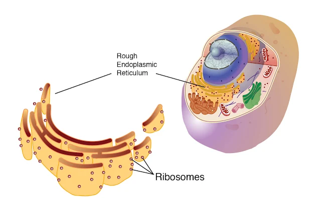
ENDOPLASMIC RETICULUM:  The endoplasmic reticulum (ER) is an extensive network of membranous tubules that extend from the nuclear membrane to the cell membrane. Rough ER has numerous ribosomes on its surface, whereas smooth ER has no ribosomes. As a network of interconnected tunnels, the ER is a passage way for the transport of the materials necessary for cell function within the cell. These include proteins synthesized by the ribosomes on the rough ER, and lipids synthesized by the smooth ER. 

RIBOSOMES:  Ribosomes are very small structures made of protein and ribosomal RNA. Some are found on the surface of rough ER, while others float freely within the cytoplasm. Ribosomes are the site of protein synthesis. The proteins produced may be structural proteins such as collagen in the skin, enzymes, or hormones such as insulin that regulate cellular processes. These proteins may function within the cell or be secreted from the cell to be used elsewhere in the body. Our protein molecules are subject to damage, and some cellular proteins, especially regulatory proteins, may be needed just for a very short time. All such proteins must be destroyed, and this is the function of proteasomes. 

PROTEOSOMES:  A proteasome is a barrel-shaped organelle made of enzymes that cut protein molecules apart (protease enzymes). Proteins that are to be destroyed, that is, those no longer needed or those that are damaged or misfolded, are tagged by a protein called ubiquitin (sort of a cellular mop or broom) and carried into a proteasome. The protein is snipped into peptides or amino acids, which may be used again for protein synthesis on ribosomes. Proteasomes are particularly important during cell division and during embryonic development, when great changes are taking place very rapidly as cells become specialized. Many of our cells have secretory functions, that is, they produce specific products to be used elsewhere in tissues. 

GOLGI APPARATUS:  Secretion is one task of the Golgi apparatus, a series of flat, membranous sacs, somewhat like a stack of saucers. Carbohydrates are synthesized within the Golgi apparatus, and are packaged, along with other materials, for secretion from the cell. Proteins from the ribosomes or lipids from the smooth endoplasmic reticulum may also be secreted in this way. To secrete a substance, small sacs of the Golgi membrane break off and fuse with the cell membrane, releasing the substance to the exterior of the cell. This is exocytosis, exo meaning “to go out” of the cell. 

MITOCHONDRIA:  Mitochondria are oval or spherical organelles bounded by a double membrane. The inner membrane has folds called cristae. Within the mitochondria, the aerobic (oxygen-requiring) reactions of cell respiration take place. Therefore, mitochondria are the site of ATP (and hence energy) production. Cells that require large amounts of ATP, such as muscle cells, have many mitochondria to meet their need for energy. Mitochondria contain their own genes in a single DNA molecule and duplicate themselves when a cell divides. An individual’s mitochondrial DNA (mDNA) is of maternal origin, that is, from the mitochondria that were present in the ovum, or egg cell, which was then fertilized by a sperm cell. The mitochondria of the sperm cell usually do not enter the ovum during fertilization, because they are not found in the head of the sperm with the chromosomes.

LYSOSOMES:  Lysosomes are single-membrane structures that contain digestive enzymes. When certain white blood cells engulf bacteria, the bacteria are digested and destroyed by these lysosomal enzymes. Worn-out cell parts and dead cells are also digested by these enzymes. This is a beneficial process, and is necessary before tissue repair can begin. But it does have a disadvantage in that lysosomal digestion contributes to inflammation in damaged tissues. An excess of inflammation can start a vicious cycle, actually a positive feedback mechanism, that results in extensive tissue damage. Many of our cells are capable of dividing, or reproducing, themselves. 

CENTRIOLES:  Centrioles are a pair of rodshaped structures perpendicular to one another, located just outside the nucleus. Their function is to organize the spindle fibers during cell division. The spindle fibers are contracting proteins that pull the two sets of chromosomes apart, toward the ends of the original cell as it divides into two new cells. Each new cell then has a full set of chromosomes. 

CILIA AND FLAGELLA:  Cilia and flagella are mobile thread-like projections through the cell membrane; each is anchored by a basal body just within the membrane. Cilia are usually shorter than flagella, and an individual cell has many of them on its free surface. The cilia of a cell beat in unison and sweep materials across the cell surface. Cells lining the fallopian tubes, for example, have cilia to sweep the egg cell toward the uterus. The only human cell with a flagellum is the sperm cell. The flagellum provides motility, or movement, for the sperm cell. 

MICROVILLI:  Microvilli are folds of the cell membrane on the free surface of a cell.


RELATED;

1. CELLULAR METABOLISM  

2.  STUCTURE OF A CELL MEMBRANE  

3.  ENZYMES

4.  PROTEINS

5.  BODY TISSUES

REFERENCES


November 01, 2020

PHYSICAL AND CHEMICAL BARRIERS TO INFECTION


INTRODUCTION:  The skin:  The squamous epithelium of the skin is the first line of defense against microorganisms encountered in the outside world. As keratinized epithelial surface cells desquamate, the skin maintains its protective barrier by generating new epithelial cells beneath the surface. The skin is also bathed with oils and moisture from the sebaceous and sweat glands. These secretions contain fatty acids that inhibit bacterial growth. Poor vascular supply to the skin may result in skin breakdown and increased susceptibility to infection. For example, chronically debilitated or bedridden patients may suffer from presacral pressure ulcers as a result of constant compression of this dependent body area; these ulcers allow direct entry of skin and enteric bacteria into previously sterile sites and predispose patients to severe infections.

The mucus membranes:  The mucous membranes also provide a physical barrier to microbial invasion. The mucous membranes of the mouth, pharynx, esophagus, and lower urinary tract are composed of several layers of epithelial cells, whereas those of the lower respiratory tract, the GI tract, and the upper urinary tract are delicate single layers of epithelial cells. These membranes are covered by a protective layer of mucus, which traps foreign particles and prevents them from accessing the lining of epithelial cells. Because mucus is hydrophilic, many substances produced by the body easily diffuse to the surface, including enzymes with antimicrobial activity such as lysozyme and peroxidase.

RELATED;

MOST FREQUENTLY READ