Showing posts with label Medicine and surgery. Show all posts

Saturday, March 25, 2023

NORMAL LABOR AND VAGINAL DELIVERY

INTRODUCTION: Labor and birth are natural human processes and for most women, minimal interventions are required. Uncomplicated labor occurs at term which is taken to be completion of 38 weeks of gestation, with a single fetus in a vertex presentation, with labor initiated by spontaneous, effective contractions, and with birth completed within 24 hours.

BEGINNING OF LABOR: Initiation of labor is a complex process involving a combination of factors that work in conjunction to stimulate myometrial activity and in turn initiate the onset of labor. These factors may include oxytocin release from the posterior pituitary, uterine distention or stretching, increasing uterine pressure due to the term fetus, increased maternal prostaglandin and fetal cortisol levels, placental aging, and changes in estrogen and progesterone ratios. The exact mechanism(s) that initiate spontaneous labor have been researched but are not completely understood.

TRUE VERSUS FALSE LABOR: Premonitory signs of labor such as lightening, urinary frequency, a change in vaginal discharge including bloody show, the loss of the mucus plug, and irregular contractions are often reported before true labor begins. True labor is distinguished from false labor by contractions that become progressively more frequent and regular, discomfort beginning in the back and radiating toward the abdomen causing cervical dilation, cervical effacement, and fetal descent. Duration of labor will depends on fetal presentation, fetal size, position of the fetus, and a multitude of factors including pelvic structure, the woman’s body mass, and birthing position.

STAGES OF LABOR: Labor is divided into four stages; 

Stage 1: The first stage of labor, which is cervical dilation to 10 cm, is divided into latent (0-3 cm dilation), active (4-7 cm dilation), and transitional (8-10 cm dilation) phases.

Stage 2: Stage two is measured from compete dilation to the birth of the baby and may have a latent or passive phase.

Stage 3: Stage three is from the birth of the baby to the expulsion of the placenta.

Stage 4: Stage four is the immediate postpartum recovery phase occurring from the delivery of the placenta and encompassing the first 2 hr postdelivery.

RELATED;

1.  PRETERM LABOR AND BIRTH

2.  DRUGS USED DURING LABOR AND DELIVERY

3.  ECTOPIC PREGNANCY

4.  PARTURITION AND LABOR

REFERENCES

Friday, October 28, 2022

FETAL CIRCULATION


INTRODUCTION: The fetus depends upon the mother for oxygen and nutrients and for the removal of carbon dioxide an other waste products. The site of exchange between fetus and mother is the placenta, which contains fetal and maternal blood vessels that are very close to one another.

THE FETAL-MATERNAL BLOOD: The blood of the fetus does not mix with the blood of the mother; substances are exchanged by diffusion and active transport mechanisms. The fetus is connected to the placenta by the umbilical cord, which contains two umbilical arteries and one umbilical vein.

THE UMBILICAL CODE: The umbilical arteries are branches of the fetal internal iliac arteries; they carry blood from the fetus to the placenta. In the placenta, carbon dioxide and waste products in the fetal blood enter maternal circulation, and oxygen and nutrients from the mother’s blood enter fetal circulation. The umbilical vein carries this oxygenated blood from the placenta to the fetus. Within the body of the fetus, the umbilical vein branches: One branch takes some blood to the fetal liver, but most of the blood passes through the ductus venosus to the inferior vena cava, to the right atrium.

NEONATAL CIRCULATION: After birth, when the umbilical cord is cut, the remnants of these fetal vessels constrict and become nonfunctional. The other modifications of fetal circulation concern the fetal heart and. Because the fetal lungs are deflated and do not provide for gas exchange, blood is shunted away from the lungs and to the body. The foramen ovale is an opening in the interatrial septum that permits some blood to flow from the right atrium to the left atrium, not, as usual, to the right ventricle. The blood that does enter the right ventricle is pumped into the pulmonary artery. The ductus arteriosus is a short vessel that diverts most of the blood in the pulmonary artery to the aorta, to the body. Both the foramen ovale and the ductus arteriosus permit blood to bypass the fetal lungs. Just after birth, the baby breathes and expands its lungs, which pulls more blood into the pulmonary circulation. More blood then returns to the left atrium, and a flap on the left side of the foramen ovale is closed. The ductus arteriosus constricts, probably in response to the higher oxygen content of the blood, and pulmonary circulation becomes fully functional within a few days.


RELATED;

1.  FETAL DIAGNOSIS

2.  HEMORRHAGIC DISEASE OF THE NEW BORN

3.  ANATOMY AND PHYSIOLOGY

REFERENCES

Sunday, September 25, 2022

ALLERGY


INTRODUCTION: An allergy is a hypersensitivity to a particular foreign antigen, called an allergen. Allergens include plant pollens, foods, chemicals in cosmetics, antibiotics such as penicillin, dust, and mold spores. Such allergens are not themselves harmful. Most people, for example, can inhale pollen, eat peanuts, or take penicillin with no ill effects.

HYPERSENSITIVITY: Hypersensitivity means that the immune system overresponds to the allergen, and produces tissue damage by doing so. Allergic responses are characterized by the production of IgE antibodies, which bond to mast cells. Mast cells are specialized connective tissue cells and are numerous in the connective tissue of the skin and mucous membranes.

INFLAMMATORY MEDIATORS: Chemicals in mast cells include histamine and leukotrienes, which are released by the bonding of IgE antibodies or when tissue damage occurs. These chemicals contribute to the process of inflammation by increasing the permeability of capillaries and venules. Tissue fluid collects and more WBCs are brought to the damaged area. In an allergic reaction, the effects of inflammatory chemicals create symptoms such as watery eyes and runny nose (hay fever) or the more serious wheezing and difficult breathing that characterize asthma. Several medications are available to counteract these effects.

ANAPHYLACTIC SHOCK: Anaphylactic shock is an extreme allergic response that may be elicited by exposure to penicillin or insect venoms. On the first exposure, the person becomes highly sensitized to the foreign antigen. On the second exposure, histamine is released from mast cells throughout the body and causes a drastic decrease in blood volume. The resulting drop in blood pressure may be fatal in only a few minutes. People who know they are allergic to bee stings, for example, may obtain a self-contained syringe of epinephrine to carry with them. Epinephrine can delay the progression of anaphylactic shock long enough for the person to seek medical attention.


RELATED;

1.  IMMUNOGLOBULINS  

2.  IMMUNISATION

3.  MEDICAL CONDITIONS

REFERENCES

Wednesday, August 03, 2022

NAUSEA AND VOMITING


INTRODUCTION: Nausea is an unpleasant, subjective sensation that is accompanied by weakness, diaphoresis, and hyperproduction of saliva. It is sometimes accompanied by dizziness. Intense nausea often leads to vomiting, or emesis.

PATHOPHYSIOLOGY: Vomiting is a defense mechanism used by the body to rid itself of toxic substances. Vomiting is a reflex primarily controlled by the vomiting center of the medulla of the brain, which receives sensory signals from the digestive tract, the inner ear, and the chemoreceptor trigger zone (CTZ) in the cerebral cortex. Interestingly, the CTZ is not protected by the blood–brain barrier, as is the vast majority of the brain; thus, these neurons can directly sense the presence of toxic substances in the blood. Once the vomiting reflex is triggered, wavelike contractions of the stomach quickly propel its contents upward and out of the body.

OBJECTIVES OF THE TREATMENT: The treatment outcomes for nausea or vomiting focus on removal of the cause, whenever feasible.

CAUSES OF NAUSEA AND VOMITING: Nausea and vomiting are common symptoms associated with a wide variety of conditions such as GI infections, food poisoning, nervousness, emotional imbalances, motion sickness, and extreme pain. Other conditions that promote nausea and vomiting are general anesthetics, migraine headache, trauma to the head or abdominal organs, inner ear disorders, and diabetes. Psychological factors play a significant role, as patients often become nauseated during periods of extreme stress or when confronted with unpleasant sights, smells, or sounds. The nausea and vomiting experienced by women during the first trimester of pregnancy is referred to as morning sickness. If this condition becomes acute, with continual vomiting, it may lead to hyperemesis gravidarum, a situation in which the health and safety of the mother and developing baby can become compromised. Pharmacotherapy is initiated after other antinausea measures have proved ineffective. Nausea and vomiting are the most frequently listed adverse effects for oral medications.

CONSEQUENCES OF NAUSEA AND VOMITING: Nausea and vomiting is a common reason for patients’ lack of adherence to the therapeutic regimen and for discontinuation of drug therapy. When large amounts of fluids are vomited, dehydration and significant weight loss may occur. Because the contents lost from the stomach are strongly acidic, vomiting may cause a change in the pH of the blood, resulting in metabolic alkalosis. With excessive loss, severe acid–base disturbances can lead to vascular collapse, resulting in death if medical intervention is not initiated. Dehydration is especially dangerous for infants, small children, and older adults and is evidenced by dry mouth, sticky saliva, and reduced urine output that is dark yellow-orange to brown.


RELATED;

1.  ANTIEMETIC AGENTS  

2.  THE ENTERIC NERVOUS SYSTEM

3.  MEDICAL CONDTIONS

REFERENCES

Saturday, July 30, 2022

JAUNDICE


Objectives this article:  By the end of this article, the learner will be able to; 
1.  understand the cause of the yellow color that appear in mucous membranes.
2.  Explain the role of the liver in elimination of bilirubin

INTRODUCTION: Jaundice is not a disease, but rather a sign caused by excessive accumulation of bilirubin in the blood. Because one of the liver’s many functions is the excretion of bilirubin, jaundice may be a sign of liver disease such as hepatitis or cirrhosis. Hepatitis

This may be called hepatic jaundice, because the problem is with the liver. Other types of jaundice are prehepatic jaundice and posthepatic jaundice: The name of each tells us where the problem is. Recall that bilirubin is the waste product formed from the heme portion of the hemoglobin of old RBCs. Hemoglobin: The human red blood cells

PATHOPHYSIOLOGY: Prehepatic jaundice means that the problem is “before” the liver; that is, hemolysis of RBCs is taking place at a more rapid rate. Rapid hemolysis is characteristic of sickle cell anemia, malaria, and Rh disease of the newborn; these are hemolytic anemias. Sickle cell anaemia: Rh disease of the newborn

As excessive numbers of RBCs are destroyed, bilirubin is formed at a faster rate than the liver can excrete it. The bilirubin that the liver cannot excrete remains in the blood and causes jaundice. Another name for this type is hemolytic jaundice.

Posthepatic jaundice means that the problem is “after” the liver. The liver excretes bilirubin into bile, which is stored in the gallbladder and then moved to the small intestine. If the bile ducts are obstructed, perhaps by gallstones or inflammation of the gallbladder, bile cannot pass to the small intestine and backs up in the liver. Bilirubin may then be reabsorbed back into the blood and cause jaundice. Another name for this type is obstructive jaundice.

RELATED;

1.  THE CYTOCHROME P450 ENZYME SYSTEM  

2.  CATALASE

3.  FUNCTIONS OF THE LIVER

4.  MEDICAL CONDITIONS

REFERENCES

Saturday, May 28, 2022

ULTERED LEVEL OF CONSCIOUSNESS


INTRODUCTION:  Unconsciousness also known as altered Level Of Consciousness (ALOC) is a condition in which the patient is unresponsive to and unaware of environmental stimuli, usually for a short duration. Coma is a clinical state which is an unarousable, unresponsive condition in which the patient is unaware of self or the environment for prolonged periods (days to months, or even years).  Akinetic mutism is a state of unresponsiveness to the environment in which the patient makes no voluntary movement. A persistent vegetative state is one in which the unresponsive patient resumes sleep–wake cycles after coma but is devoid of cognitive or affective mental function. Locked-in syndrome results from a lesion affecting the pons and results in paralysis and the inability to speak, but vertical eye movements and lid elevation remain intact and are used to indicate responsiveness.

CAUSES OF ALOC:  The causes of unconsciousness may be neurologic for example resulting from head injury or stroke, toxicologic such as following drug overdose, alcohol intoxication, or metabolic for example following hepatic or renal failure, diabetic ketoacidosis among others.

ASSESSMENT AND DIAGNOSTIC METHODS:  Neurologic examination (CT, MRI, positron emission tomography [PET], electroencephalography [EEG], single photon emission CT [SPECT]) to identify cause of loss of consciousness. Laboratory tests include; analysis of blood glucose, electrolytes, serum ammonia, and liver function tests; blood urea nitrogen (BUN) levels; serum osmolality; calcium level; and partial thromboplastin and prothrombin times.  Other studies may be used to evaluate serum ketones, alcohol and drug concentrations, and arterial blood gases.

MEDICAL MANAGEMENT:  The first priority is a patent and secure airway (intubation or tracheostomy). Then circulatory status (carotid pulse, heart rate and impulse, blood pressure) is assessed and adequate oxygenation maintained. An IV line is established to maintain fluid balance status, and nutritional support is provided (feeding tube or gastrostomy).  Neurologic care is based on specific pathology. Other measures include drug therapy and measures to prevent complications.


RELATED;

1.  The central nervous system

2.  Meningitis

3.  Hypoglycemia

4.  Parkinson's disease

5.  Medicine and surgery

REFERENCES

Monday, May 23, 2022

INFLAMMATORY BOWEL DISEASE


CLINICAL PRESENTATION:  Inflammatory bowel disease is distinguished from infectious entities by exclusion and by chronicity (months; not days or weeks). Patients often experience recurrent episodes of mucopurulent, bloody diarrhea characterized by lack of positive cultures for known microbial pathogens and failure to respond to antibiotics alone. Antibiotics:  Microbial pathogens

Because inflammatory bowel disease is characterized by exacerbations and remissions, favorable responses to therapy may be difficult to distinguish from spontaneous remissions occurring as part of the natural history of the disease.

ETIOLOGY:  Triggers for the development of IBD remain elusive. However, epidemiologic studies suggest that smoking and gastrointestinal infections are possible contributory factors. There are two forms of chronic inflammatory bowel disease: Crohn’s disease, which is transmural and granulomatous in character, occurring anywhere along the GI tract; and ulcerative colitis, which is superficial and limited to the colonic mucosa.  Ulcerative colitis

The exact causes of inflammatory bowel disease are unknown despite progress in understanding its pathogenesis.

PATHOLOGY & PATHOGENESIS:  Genetic risk and environmental factors are recognized as two key elements in the pathogenesis of inflammatory bowel disease. An explosion of newly recognized susceptibility genes for both Crohn disease and ulcerative colitis have been discovered through genome-wide associations. These studies evaluated thousands of single nucleotide polymorphisms (SNPs) in thousands of patients with inflammatory bowel disease and compared them with SNPs from thousands of people without the disease (controls). These studies found abnormalities in several categories of susceptibility genes in patients with inflammatory bowel disease. These included modulators of immune function, autophagy, and epithelial function that participate in the interaction of host and microorganism. Importantly, the relative risk of most of these susceptibility genes is low (most have a 20–30% increase in relative risk of developing disease).  Therefore, most people who harbor risk alleles for inflammatory bowel disease do not develop disease.  Genetic factors are clearly not the sole contributor to inflammatory bowel disease.  Many environmental factors have been found to contribute to the development of Crohn’s disease, including pathogenic microorganisms (bacteria and viruses), the repertoire of indigenous intestinal microbes (the microbiota), dietary factors, smoking, defective immune responses, and psychosocial factors.


RELATED;

1.  CONSTIPATION

2.  ULCERATIVE COLITIS

3.  MEDICAL CONDITIONS

REFERENCES

Sunday, May 22, 2022

DIARRHEA


INTRODUCTION:  Diarrhea is a condition defined by an increased frequency of bowel movements that is to say, more than three per day, increased amount of stool, and altered consistency which is usually referred to as liquid stool. It is usually associated with urgency, perianal discomfort, incontinence, or a combination of these factors. Diarrhea can result from any condition that causes increased intestinal secretions, decreased mucosal absorption, or altered motility.

TYPES OF DIARRHEA:  Types of diarrhea include secretory, osmotic, malabsorptive, infectious, and exudative. It can be acute where it can be self-limiting and often associated with infection, or chronic which persists for a long period and may return sporadically. It can be caused by certain medications, tube feeding formulas, metabolic and endocrine disorders, and viral and bacterial infections. Other causes are nutritional and malabsorptive disorders, anal sphincter deficit, Zollinger–Ellison syndrome, paralytic ileus, acquired immunodeficiency syndrome (AIDS), and intestinal obstruction. Intestinal obstruction:  AIDS

CLINICAL MANIFESTATIONS:  Increased frequency and fluid content of stool.  Abdominal cramps, distention, intestinal rumbling, anorexia, and thirst.  Painful spasmodic contractions of the anus and ineffectual straining (tenesmus) with each defecation.  Other symptoms, depending on the cause and severity and related to dehydration and fluid and electrolyte imbalances, include the following: Watery stools, which may indicate small bowel disease.  Inflammatory bowel disease  

Loose, semisolid stools, which are associated with disorders of the large bowel.  Voluminous greasy stools, which suggest intestinal malabsorption.  Blood, mucus, and pus in the stools, which denote inflammatory enteritis or colitis.  Oil droplets on the toilet water, which are diagnostic of pancreatic insufficiency.  Nocturnal diarrhea, which may be a manifestation of diabetic neuropathy.

COMPLICATIONS:  Complications of diarrhea include cardiac dysrhythmias due to fluid and electrolyte (potassium) imbalance, urinary output less than 30 mL/h, muscle weakness, paresthesia, hypotension, anorexia, drowsiness, skin care issues related to irritant dermatitis, and death if imbalances become severe.

ASSESSMENT AND DIAGNOSTIC FINDINGS:  When the cause is not obvious: complete blood cell count; serum chemistries; urinalysis; routine stool examination; and stool examinations for infectious or parasitic organisms, bacterial toxins, blood, fat, electrolytes, and white blood cells. Endoscopy or barium enema may assist in identifying the cause. 

MEDICAL MANAGEMENT:  Primary medical management is directed at controlling symptoms, preventing complications, and eliminating or treating the underlying disease.  Certain medications such as, antibiotics, anti-inflammatory agents and antidiarrheals like, loperamide, diphenoxylate may reduce the severity of diarrhea and the disease.  Increase oral fluids; oral glucose and electrolyte solution may be prescribed.  Antimicrobials are prescribed when the infectious agent has been identified or diarrhea is severe. IV therapy is used for rapid hydration in very young or elderly patients.

RELATED;

1.  CONSTIPATION

2.  ULCERATIVE COLITIS

3.  INFLAMMATORY BOWEL DISEASE

4.  PEPTIC ULCER DISEASE

REFERENCES

Saturday, May 21, 2022

MEGALOBLASTIC ANEMIA


INTRODUCTION:  In the anemias caused by deficiencies of vitamin B12 or folic acid, identical bone marrow and peripheral blood changes occur because both vitamins are essential for normal DNA synthesis.

PATHOPHYSIOLOGY:  Folic Acid Deficiency: Folic acid is stored as compounds referred to as folates. The folate stores in the body are much smaller than those of vitamin B12, and they are quickly depleted when the dietary intake of folate is deficient ( usually within 4 months). Folate deficiency occurs in people who rarely eat uncooked vegetables. Alcohol increases folic acid requirements; folic acid requirements are also increased in patients with chronic hemolytic anemias and in women who are pregnant. Some patients with malabsorptive diseases of the small bowel may as well not absorb folic acid normally.

Vitamin B12 Deficiency: A deficiency of vitamin B12 can occur in several ways. Inadequate dietary intake is rare but can develop in strict vegetarians who consume no meat or dairy products.  Faulty absorption from the GI tract is more common, as with conditions such as Crohn’s disease or after ileal resection or gastrectomy. Another cause is the absence of intrinsic factor. A deficiency may also occur if disease involving the ileum or pancreas impairs absorption.  The body normally has large stores of vitamin B12, so years may pass before the deficiency results in anemia.

CLINICAL MANIFESTATIONS:  Symptoms of folic acid and vitamin B12 deficiencies are similar, and the two anemias may coexist. Symptoms are progressive, although the course of illness may be marked by spontaneous partial remissions and exacerbations.  Gradual development of signs of anemia (weakness, listlessness, and fatigue).  Possible development of a smooth, sore, red tongue and mild diarrhea (pernicious anemia).  Mild jaundice, vitiligo.  Confusion may occur; more often, paresthesias in the extremities and difficulty keeping balance; loss of position sense.  Lack of neurologic manifestations with folic acid deficiency alone.  Without treatment, patients die, usually as a result of heart failure secondary to anemia.  Heart failure

ASSESSMENT AND DIAGNOSTIC FINDINGS:  Schilling test (primary diagnostic tool).  Complete blood cell count (Hgb value as low as 4 to 5 g/dL, WBC count 2,000 to 3,000 per ml of blood, platelet count fewer than 50,000 per ml of blood; very high MCV).  Serum levels of folate and vitamin B12 (folic acid deficiency and deficient vitamin B12)

MEDICAL MANAGEMENT: Folic Acid Deficiency:  Increase intake of folic acid in patient’s diet and administer 1 mg folic acid daily.  Administer IM folic acid for malabsorption syndromes. Prescribe additional supplements as necessary, because the amount in multivitamins may be inadequate to fully replace deficient body stores.  Prescribe folic acid for patients with alcoholism as long as they continue to consume alcohol.

Vitamin B12 Deficiency:  Provide vitamin B12 replacement: Vegetarians can prevent or treat deficiency with oral supplements with vitamins or fortified soy milk; when the deficiency is due to the more common defect in absorption or the absence of intrinsic factor, replacement is by monthly IM injections of vitamin B12.  A small amount of an oral dose of vitamin B12 can be absorbed by passive diffusion, even in the absence of intrinsic factor, but large doses (2 mg/day) are required if vitamin B12 is to be replaced orally.  To prevent recurrence of pernicious anemia, vitamin B12 therapy must be continued for life.

RELATED;

1.  Anemia

2.  Iron deficiency anemia

3.  Medical conditions

REFERENCES

Tuesday, March 29, 2022

PNEUMONIA

INTRODUCTION: Pneumonia is an inflammation of the lung parenchyma caused by various microorganisms, including bacteria, mycobacteria, fungi, and viruses. Pneumonias are classified as community acquired pneumonia (CAP), hospital-acquired (nosocomial) pneumonia (HAP), pneumonia in the immunocompromised host, and aspiration pneumonia. There is overlap in how specific pneumonias are classified, because they may occur in differing settings. Those at risk for pneumonia often have chronic underlying disorders, severe acute illness, a suppressed immune system from disease or medications, immobility, and other factors that interfere with normal lung protective mechanisms. The elderly are also at high risk.

PATHOPHYSIOLOGY: An inflammatory reaction can occur in the alveoli, producing an exudate that interferes with the diffusion of oxygen and carbon dioxide; Oxygen; Carbon dioxide  bronchospasm may also occur if the patient has reactive airway disease. Bronchopneumonia, the most common form, is distributed in a patchy fashion extending from the bronchi to surrounding lung parenchyma. Lobar pneumonia is the term used if a substantial part of one or more lobes is involved. Pneumonias are caused by a variety of microbial agents in the various settings. Common organisms include Pseudomonas aeruginosa and Klebsiella species; Staphylococcusaureus; and entericGram-negative bacilli, and viruses.

CLINICAL MANIFESTATIONS: Clinical features vary depending on the causative organism and the patient’s disease. Sudden chills and rapidly rising fever of a temperature raising from 38.50C to 40.50C. Pleuritic chest pain aggravated by respiration and coughing. Severely ill patient has marked tachypnea (25 to 45 breaths/min) and dyspnea; orthopnea when not propped up. Pulse rapid and bounding; may increase 10 beats/min per degree of temperature elevation. A relative bradycardia for the amount of fever suggests viral infection, mycoplasma infection, or infection with a Legionella organism.

OTHER SIGNS: upper respiratory tract infection, headache, low-grade fever, pleuritic pain, myalgia, rash, and pharyngitis; after a few days, mucoid or mucopurulent sputum is expectorated.

SEVERE PNEUMONIA: flushed cheeks; lips and nail beds demonstrating central cyanosis. Sputum purulent, rusty, blood-tinged, viscous, or green depending on etiologic agent. Appetite is poor, and the patient is diaphoretic and tires easily. Signs and symptoms of pneumonia may also depend on a patient’s underlying condition for example, different signs occur in patients with conditions such as cancer, and in those who are undergoing treatment with immunosuppressants, which decrease the resistance to infection.

ASSESSMENT AND DIAGNOSTIC METHODS: Primarily history, physical examination. Chest x-rays, blood and sputum cultures, Gram stain.

MEDICAL MANAGEMENT: Antibiotics are prescribed on the basis of Gram stain results and antibiotic guidelines including resistance patterns, risk factors, etiology must be considered. Combination therapy may also be used. Supportive treatment includes hydration, antipyretics, antitussive medications, antihistamines, or nasal decongestants. Bed rest is recommended until infection shows signs of clearing. Oxygen therapy is given for hypoxemia. Respiratory support includes high inspiratory oxygen concentrations, endotracheal intubation, and mechanical ventilation. For groups at high risk for CAP, pneumococcal vaccination is advised.


RELATED;

1.  TUBERCULOSIS

2.  ASTHMA

3.  ACUTE RESPIRATORY DISTRESS SYNDROME

4.  MEDICAL CONDITIONS

REFERENCES

Saturday, February 12, 2022

IRON DEFICIENCY ANEMIA


Introduction: Iron-deficiency anemia typically results when the intake of dietary iron is inadequate for hemoglobin synthesis. Iron deficiency anemia is the most common type of anemia in all age groups, and it is the most common anemia in the world. The most common cause of iron-deficiency anemia in men and postmenopausal women is bleeding from ulcers, gastritis, inflammatory bowel disease, or GI tumors. PEPTIC ULCERS,  The most common causes of iron-deficiency anemia in premenopausal women are menorrhagia that is to say, excessive menstrual bleeding and pregnancy with inadequate iron supplementation. Patients with chronic alcoholism often have chronic blood loss from the GI tract, which causes iron loss and eventual anemia. Other causes include iron malabsorption, as is seen after gastrectomy or with celiac disease.

Clinical Manifestations: Symptoms of anemia. Symptoms in more severe or prolonged cases: smooth, sore tongue; brittle and ridged nails; angular cheilosis (mouth ulceration)

Assessment and Diagnostic Methods: Bone marrow aspiration. Laboratory values, including serum ferritin levels (indicates iron stores), blood cell count (hemoglobin, hematocrit, RBC count, MCV), serum iron level, and total iron-binding capacity

Medical Management: Search for the cause, which may be a curable GI cancer or uterine fibroids. Test stool specimens for occult blood. People aged 50 years or older should have periodic colonoscopy, endoscopy, or x-ray examination of the GI tract to detect ulcerations, gastritis, polyps, or cancer. Administer prescribed iron preparations (oral, intramuscular [IM], or IV). Have patient continue iron preparations for 6 to 12 months.

RELATED;

1. BLOOD AND ITS COMPONENTS

2. BIOMOLECULES AND CHEMICALS OF LIFE

3.  OTHER TYPES OF ANEMIA

REFERENCES

Saturday, July 31, 2021

AUTOIMMUNE DISEASES


INTRODUCTION:  Autoimmunity is the body's malfunctioning of destroying it's own tissues.  In this natural phenomenon, the immune system tend to mistakenly recognize body tissues as foreign and it wedges attack against it.  The root causes of this destructive auto process is as a result of body's previous exposure to molecules that resemble the body's human leukocyte antigen discussed in some of our previous chapters. In our discussion here, let us look at the most common autoimmune disease that we encounter and their common presentation.  If you did not follow us on the discussion on the pathophysiology of autoimmunity, click on the link below to get the details about the condition.  The human leukocyte antigen and development of autoimmunity


HASHIMOTO’S THYROIDITIS:  This was the first disease that satisfied Witebsky’s criteria for an autoimmune disease.  Hashimoto’s disease is characterised physiologically by a deficiency in thyroid hormone and anatomically by an enlarged thyroid gland infilterated with plasma cells and lymphocytes.


THROMBOCYTOPENIC PURPURA:  Thrombocytopenic purpura is an illness characterised by thrombocytopenia and the appearance of purpuric or petechial haemorrhages in the skin and tissues.  If you have not been following us click here to read about thrombocytopenia.  In infants, this condition can arise through allo-immunisation in much the same way that erythroblastosisfetalis develops.  An autoimmune form of thrombocytopenic purpura is seen in adults as well in which in most cases, it is drug-induced.  Invariably the afflicted person is on a continued drug regimen of some sort.  The offending drug may be aspirin, sulfonamide, quinine, antihistaminics, and many others.


MYASTHENIA GRAVIS:  This is a disease in which a gradual progressive weakness of striated muscle is a prominent external sign and which becomes so severe that even eating is laborious.  Myasthenia gravis patients exhibit a number of immunologic aberrations. Antinuclear antibodies, rheumatoid factors and antibodies that react with striated muscles are frequently detected.  Commonest immunopathological feature is presence of antibody to acetylcholine receptor.


MULTIPLE SCLEROSIS:  Partial loss of vision, nystagmus, facial palsy and muscular incoordination are a few of the varied symptoms of multiple sclerosis.  Remissions and exacerbations are characteristics of this disease. The major pathologic feature is an inflammatory lesion of the myelin in the central nervous system.

GLOMERULONEPHRITIS:  There are three forms of immune diseases that involve the glomerulus.  One is associated with antecedent group A streptococcal infection, second is involved with heterologous antibodies versus glomerular basement membrane antigens and third is based on immune complex formation with foreign antigens, as in serum sickness or alloantigens, as in SLE.

RHEUMATOID ARTHRITIS:  It is inflammatory disease of joints and connective tissue, amyloid deposition of tissues and permanent deformity of the joints may result.  An autoantibody seen in this condition is an agglutinating agent known as rheumatoid factor (RF) which is a 19S Ig compatible in all respects with IgM.

GRAVES’ DISEASE:  This disease is a form of hyperthyroidism and more common than Hashimoto’s disease.  It is now believed to be caused by a long acting thyroid stimulator (LATS) an immunoglobulin found in the sera of majority of patients with Graves’ disease.  The LATS autoantibody is a type of enhancing antibody that operates at the cellular level, much like the thyroid stimulating hormone, to stimulate thyroid hormone release and thyrotoxicosis.  

RELATED;

1.  Thrombocytopenia

2.  Specific immunity

3.  Passive immunisation

4.  Thrombocytopenia

5.  Hemorrhagic disease of the new born


Saturday, October 31, 2020

PRURITUS

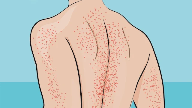
INTRODUCTION: Pruritus also known as itching is one of the most common dermatologic complaints. Scratching the itchy area causes the inflamed cells and nerve endings to release histamine, which produces more pruritus and, in turn, a vicious itch–scratch cycle. Scratching can result in altered skin integrity with excoriation, redness, raised areas or what one may call wheals, infection, or changes in pigmentation. Although pruritus usually is due to primary skin disease, it may also reflect systemic internal disease, such as diabetes mellitus; renal, hepatic, thyroid, or blood disorders; or cancer. Pruritus may be caused by certain oral medications including but not limited to; aspirin, antibiotics, hormones and opioids. Among other causes we may have, contact with irritating agents such as soaps, chemicals, or prickly heat. It may also be a side effect of radiation therapy, a reaction to chemotherapy, or a symptom of infection. It may occur in elderly patients as a result of dry skin as well. It may also be caused by psychological factors such as emotional stress.

CLINICAL MANIFESTATIONS: Itching and scratching, often more severe at night. Excoriations, redness, raised areas on the skin, as a result of scratching. Infections or changes in pigmentation. Debilitating itching, in severe cases.

MEDICAL MANAGEMENT: The cause of pruritus needs to be identified and treated. The patient is advised to avoid washing with soap and hot water. Cold compresses, ice cubes, or cool agents that contain soothing menthol and camphor may be applied. Bath oils are prescribed, except for elderly patients or those with impaired balance, who should not add oil to the bath because of the danger of slipping. Topical corticosteroids are prescribed to decrease itching. Oral antihistamines such as diphenhydramine may be used.

RELATED;

1.  SEBORRHEIC DERMATITIS

2.  BURNS

3.  WOUND HEALING

REFERENCES


Wednesday, July 22, 2020

THROMBOPHLEBITIS AND DEEP VEIN THROMBOSIS

INTRODUCTION: Thrombophlebitis is an inflammation of the walls of the veins, often accompanied by the formation of a clot. When a clot develops initially in the veins as a result of stasis or hypercoagulability, but without inflammation, the process is referred to as phlebothrombosis. Venous thrombosis can occur in any vein but is most frequent in the veins of the lower extremities than the upper extremities. Both superficial and deep veins of the legs may be affected. Damage to the lining of blood vessels creates a site for clot formation, and increased blood coagulability occurs in patients who abruptly stop taking anticoagulant medications and also occurs with oral contraceptive use and several blood dyscrasias. The danger associated with venous thrombosis is that parts of a clot can become detached and produce an embolic occlusion of the pulmonary blood vessels.

RISK FACTORS: History of varicose veins, hypercoagulation, neoplastic disease, cardiovascular disease, or recent major surgery or injury. Obesity, Advanced age, Oral contraceptive use.

CLINICAL MANIFESTATIONS: Signs and symptoms are nonspecific. Edema and swelling of the extremity resulting from obstruction of the deep veins of the leg; bilateral swelling may be difficult to detect because of lack of size difference. Skin over the affected leg may become warmer; superficial veins may become more prominent showing cordlike venous segment. Tenderness occurs later and is detected by gently palpating the leg. In some cases, signs of a pulmonary embolus are the first indication of DVT. Pulmonary embolism

Thrombus of superficial veins produces pain or tenderness, redness, and warmth in the involved area. In massive iliofemoral venous thrombosis, the entire extremity becomes massively swollen, tense, painful, and cool to touch.

ASSESSMENT AND DIAGNOSTIC METHODS: History revealing risk factors such as varicose veins or neoplastic disease. Doppler ultrasonography, duplex ultrasonography, air plethysmography, contrast phlebography (venography).

PREVENTION: Prevention is dependent on identifying risk factors for thrombus and on educating the patient about appropriate interventions.

MEDICAL MANAGEMENT: Objectives of management are to prevent the thrombus from growing and fragmenting, resolve the current thrombus, and prevent recurrence.

PHARMACOLOGIC THERAPY: Unfractionated heparin is administered for 5 days by intermittent or continuous intravenous (IV) infusion.

Dosage: Regulated by monitoring the activated partial thromboplastin time (APTT), the international normalized ratio (INR), and the platelet count. Low-molecular-weight heparin (LMWH) is given in one or two injections daily; it is more expensive than unfractionated heparin but safer. Oral anticoagulants such as, warfarin [Coumadin]) are given with heparin therapy. Thrombolytic (fibrinolytic) therapy such as, alteplase, is given within the first 3 days after acute thrombosis. Throughout therapy, PTT, prothrombin time (PT), hemoglobin and hematocrit levels, platelet count, and fibrinogen level are monitored frequently. Drug therapy is discontinued if bleeding occurs and cannot be stopped.

ENDOVASCULAR MANAGEMENT: Endovascular management is necessary for DVT when anticoagulant or thrombolytic therapy is contraindicated, the danger of pulmonary embolism is extreme, or venous drainage is so severely compromised that permanent damage to the extremity is likely. A thrombectomy may be necessary.

RELATED;

1. ARTERIOSCLEROSIS

REFERENCES


MOST FREQUENTLY READ