Showing posts with label 2024. Show all posts

August 25, 2025

ATROPINE


Therapeutic Class: Antidote for anticholinesterase poisoning

Pharmacologic Class: Muscarinic cholinergic receptor blocker.

ACTIONS AND USES:  By occupying muscarinic receptors, atropine blocks the parasympathetic actions of Ach and induces symptoms of the fight-or-flight response. Most prominent are increased heart rate, bronchodilation, decreased motility in the GI tract, mydriasis, and decreased secretions from glands.  

At therapeutic doses, atropine has no effect on nicotinic receptors in ganglia or on skeletal muscle. Although atropine has been used for centuries for a variety of purposes, its use has declined in recent decades because of the development of safer and more effective medications. Atropine may be used to treat hypermotility diseases of the GI tract such as irritable bowel syndrome, to suppress secretions during surgical procedures, to increase the heart rate in patients with bradycardia, and to dilate the pupil during eye examinations. Once widely used to cause bronchodilation in patients with asthma, atropine is now rarely prescribed for this disorder. Atropine therapy is useful for the treatment of reflexive bradycardia in infants and infantile hypertrophic pyloric stenosis (IHPS).


ADMINISTRATION ALERTS

1.  Oral and subcutaneous doses are not interchangeable.

2. Monitor blood pressure, pulse, and respirations before administration and for at least 1 hour after subcutaneous administration.

3. Pregnancy category C.


ADVERSE EFFECTS:  The side effects of atropine limit its therapeutic usefulness and are predictable extensions of its autonomic actions. Expected side effects include dry mouth, constipation, urinary retention, and an increased heart rate. Initial CNS excitement may progress to delirium and even coma.

CONTRAINDICATIONS: Atropine is contraindicated in patients with glaucoma, because the drug may increase pressure within the eye. Atropine should not be administered to patients with obstructive disorders of the GI tract, paralytic ileus, bladder neck obstruction, benign prostatic hyperplasia, myasthenia gravis, cardiac insufficiency, or acute hemorrhage.

INTERACTIONS:  Drug–Drug: Drug interactions with atropine include an increased effect with antihistamines, TCAs, quinidine, and procainamide. Atropine decreases effects of levodopa.

TREATMENT OF OVERDOSE:  Overdose may cause CNS stimulation or depression. A short-acting barbiturate or diazepam (Valium) may be administered to control convulsions. Physostigmine is an antidote for atropine poisoning that quickly reverses the coma caused by large doses of atropine.


RELATED;

1.  ACETYLCHOLINE

2.  DIAZEPAM

3.  PHARMACOLOGY AND THERAPEUTICS

REFERENCES


INFERTILITY


Definition: Infertility is defined as the absence of conception after at least 1 year of regular sexual intercourse.

Causes: Males are found to be solely responsible for 20-30% of infertility cases and these are related to issues such as, inadequate sperm count which contribute to 50% of cases.  For female infertility, about 40% of cases are due to ovulatory failure, about 40% are due to endometrial or tubal disease, about 10% are due to rarer causes such as, thyroid disease or hyperprolactinemia, and about 10% remain unexplained after full workup.

Pathophysiology of female infertility

Ovulatory Causes:  Infertility due to ovarian dysfunction can result from disorders of the hypothalamus or pituitary, resulting in inadequate gonadotropic stimulation of the ovary.  This can bring problems ranging from ovarian disorders, resulting either in inadequate secretory products or failure to ovulate; and occasionally from both types of disorder occurring at the same time. Correction of the underlying cause will often restore fertility. In many cases, the administration of exogenous gonadotropins will stimulate the ovaries to produce follicular growth. The oocytes can then be released in vivo and fertilized by intercourse or by artificial insemination.

Tubal and Pelvic Causes:  With normal follicles and reproductive neuroendocrine axis function, the major cause of infertility is an abnormality in the endometrium or fallopian tubes. Prior or ongoing pelvic infections, with adhesions or inflammation, can result in a failure of sperm or egg transport, a failure of implantation, or implantation in an inappropriate location (ectopic pregnancy).


RELATED;

1.  PELVIC INFLAMMATORY DISEASE

2.  OBSTETRICS AND GYNECOLOGY

3.  CONTRACEPTION

4.  MEDICAL CONDITIONS

REFERENCES


ALUMINUM HYDROXIDE

Therapeutic Class: Antiheartburn agent

Pharmacologic Class: Antacid

ACTIONS AND USES:  Aluminum hydroxide is an inorganic agent used alone or in combination with other antacids. Combining aluminum compounds with magnesium, increases their effectiveness and reduces the potential for constipation. Unlike calcium-based antacids that can be absorbed and cause systemic effects, aluminum compounds are minimally absorbed.  Their primary action is to neutralize stomach acid by raising the pH of the stomach contents.

Unlike H2-receptor antagonists and PPIs, aluminum antacids do not reduce the volume of acid secretion. They are most effectively used in combination with other antiulcer drugs for the symptomatic relief of heartburn due to PUD or GERD.

ADMINISTRATION ALERTS:  Administer aluminum antacids at least 2 hours before or after other drugs because absorption could be affected.  Pregnancy category C

ADVERSE EFFECTS: When taken regularly or in high doses, aluminum antacids cause constipation.

At high doses, aluminum products bind with phosphate in the GI tract and long-term use can result in phosphate depletion. Those at risk include those who are malnourished, alcoholics, and those with renal disease.

Contraindications: This drug should not be used in patients with suspected bowel obstruction.

INTERACTIONS:  Drug–Drug: Aluminum compounds should not be taken at the same time as

other medications, because they may interfere with absorption.


RELATED;

1.  PEPTIC ULCER DISEASE

2.  CONSTIPATION

3.  HISTAMINE 2 RECEPTOR BLOCKERS

4.  DIARRHEA

5.  PHARMACOLOGY AND THERAOEUTICS

REFERENCES


FETAL CIRCULATION


INTRODUCTION: The fetus depends upon the mother for oxygen and nutrients and for the removal of carbon dioxide an other waste products. The site of exchange between fetus and mother is the placenta, which contains fetal and maternal blood vessels that are very close to one another.

THE FETAL-MATERNAL BLOOD: The blood of the fetus does not mix with the blood of the mother; substances are exchanged by diffusion and active transport mechanisms. The fetus is connected to the placenta by the umbilical cord, which contains two umbilical arteries and one umbilical vein.

THE UMBILICAL CODE: The umbilical arteries are branches of the fetal internal iliac arteries; they carry blood from the fetus to the placenta. In the placenta, carbon dioxide and waste products in the fetal blood enter maternal circulation, and oxygen and nutrients from the mother’s blood enter fetal circulation. The umbilical vein carries this oxygenated blood from the placenta to the fetus. Within the body of the fetus, the umbilical vein branches: One branch takes some blood to the fetal liver, but most of the blood passes through the ductus venosus to the inferior vena cava, to the right atrium.

NEONATAL CIRCULATION: After birth, when the umbilical cord is cut, the remnants of these fetal vessels constrict and become nonfunctional. The other modifications of fetal circulation concern the fetal heart and. Because the fetal lungs are deflated and do not provide for gas exchange, blood is shunted away from the lungs and to the body. The foramen ovale is an opening in the interatrial septum that permits some blood to flow from the right atrium to the left atrium, not, as usual, to the right ventricle. The blood that does enter the right ventricle is pumped into the pulmonary artery. The ductus arteriosus is a short vessel that diverts most of the blood in the pulmonary artery to the aorta, to the body. Both the foramen ovale and the ductus arteriosus permit blood to bypass the fetal lungs. Just after birth, the baby breathes and expands its lungs, which pulls more blood into the pulmonary circulation. More blood then returns to the left atrium, and a flap on the left side of the foramen ovale is closed. The ductus arteriosus constricts, probably in response to the higher oxygen content of the blood, and pulmonary circulation becomes fully functional within a few days.


RELATED;

1.  FETAL DIAGNOSIS

2.  HEMORRHAGIC DISEASE OF THE NEW BORN

3.  ANATOMY AND PHYSIOLOGY

REFERENCES

DIAZEPAM


Introduction:  Diazepine in trade names like Valium is one of the most commonly used Over The Counter (OTC) central nervous system agents 

Therapeutic Class: Antiseizure, Sedative-hypnotic drug 

Pharmacologic Class: Benzodiazepine; GABAA receptor agonist

ACTIONS AND USES: Diazepam binds to the GABA receptor–chloride channels throughout the CNS. It produces its effects by suppressing neuronal activity in the limbic system and subsequent impulses that might be transmitted to the reticular activating system. Effects of this drug are suppression of abnormal neuronal foci that may cause seizures, calming without strong sedation, and skeletal muscle relaxation. When used orally, maximum therapeutic effects may take from 1 to 2 weeks. Tolerance may develop after about 4 weeks. When given IV, effects occur in minutes, and its anticonvulsant effects last about 20 minutes.

ADMINISTRATION ALERTS: When administering IV, monitor respirations every 5 to 15 minutes. Have airway and resuscitative equipment accessible. The drug is pregnancy category D.

ADVERSE EFFECTS: Because of tolerance and dependency, use of diazepam is reserved for short term seizure control or for status epilepticus. When given IV, hypotension, muscular weakness, tachycardia, and respiratory depression are common.

Contraindications: When administered in injectable form, this medication should be avoided under the following conditions: shock, coma, depressed vital signs, obstetrical patients, and infants less than 30 days of age. In tablet form, the medication should not be administered to infants less than 6 months of age, to patients with acute narrow-angle glaucoma or untreated open-angle glaucoma, or within 14 days of monoamine oxidase inhibitor (MAOI) therapy.

INTERACTIONS: Drug–Drug: Diazepam should not be taken with alcohol or other CNS depressants because of combined sedation effects. Other drug interactions include cimetidine, oral contraceptives, valproic acid, and metoprolol, which potentiate diazepam’s action; and levodopa and barbiturates, which decrease diazepam’s action. Diazepam increases the levels of phenytoin in the bloodstream and may cause phenytoin toxicity.


RELATED;

1.  SEDATIV-HYPNOTICS  

2.  BENZODIAZEPINES

3.  DEPRESSION

4.  PHARMACOLOGY AND THERAPEUTICS

REFERENCES

CANCER


INTRODUCTION: Cancer is a disease process that begins when an abnormal cell is transformed by the genetic mutation of the cellular DNA. The abnormal cell forms a clone and begins to proliferate abnormally, ignoring growth-regulating signals in the environment surrounding the cell. The cells acquire invasive characteristics, and changes occur in surrounding tissues. The cells infiltrate these tissues and gain access to lymph and blood vessels, which carry the cells to other areas of the body. This phenomenon is called metastasis. In otherwards the cancer spread to other parts of the body.

DESCRIPTION OF CANCER: Cancerous cells are described as malignant neoplasms and are classified and named by tissue of origin. The failure of the immune system to promptly destroy abnormal cells permits these cells to grow too large to be managed by normal immune mechanisms. Certain categories of agents or factors implicated in carcinogenesis include viruses and bacteria, physical agents, chemical agents, genetic or familial factors, dietary factors, and hormonal agents.

CLINICAL MANIFESTATIONS: Cancerous cells spread from one organ or body part to another by invasion and metastasis; therefore, manifestations are related to the system affected and degree of disruption. Generally, cancer causes anemia, weakness, weight loss, and pain which is often in late stages. Symptoms are from tissue destruction and replacement with nonfunctional cancer tissue or overproductive cancer tissue such as, bone marrow disruption and anemia or excess adrenal steroid production; pressure on surrounding structures; increased metabolic demands; and disruption of production of blood cells.

ASSESSMENT AND DIAGNOSTIC METHODS: Screening to detect early cancer usually focuses on cancers with the highest incidence or those that have improved survival rates if diagnosed early. Examples of these cancers include breast, colorectal, cervical, endometrial, testicular, skin, and oropharyngeal cancers. Patients with suspected cancer undergo extensive testing to;

1) Determine the presence and extent of tumor.

2) Identify possible spread (metastasis) of disease or invasion of other body tissues.

3) Evaluate the function of involved and uninvolved body systems and organs.

4) Obtain tissue and cells for analysis, including evaluation of tumor stage and grade.

Diagnostic tests may include tumor marker identification, genetic profiling, imaging studies (mammography, magnetic resonance imaging [MRI], computed tomography [CT], fluoroscopy, ultrasonography, endoscopy, nuclear medicine imaging, positron emission tomography [PET], PET fusion, radioimmunoconjugates), and biopsy.


RELATED;

1.  THE ORIGIN OF CANCER

2.  PATHOLOGY

REFERENCES


August 13, 2025

OXYTOCIN


INTRODUCTION: Oxytocin stimulates contraction of the uterus at the end of pregnancy and stimulates release of milk from the mammary glands. As labor begins, the cervix of the uterus is stretched, which generates sensory impulses to the hypothalamus, which in turn stimulates the posterior pituitary to release oxytocin. Oxytocin then causes strong contractions of the smooth muscle also known as ,myometrium, of the uterus to bring about delivery of the baby and the placenta.

SECRETION OF OXYTOCIN: The secretion of oxytocin is one of the few positive feedback mechanisms within the body, and the external brake or shutoff of the feedback cycle is delivery of the baby and the placenta. It has been discovered that the placenta itself secretes oxytocin at the end of gestation and in an amount far higher than that from the posterior pituitary gland. Research is continuing to determine the exact mechanism and precise role of the placenta in labor.

When a baby is breast-fed, the sucking of the baby stimulates sensory impulses from the mother’s nipple to the hypothalamus. Nerve impulses from the hypothalamus to the posterior pituitary cause the release of oxytocin, which stimulates contraction of the smooth muscle cells around the mammary ducts. This release of milk is sometimes called the “milk let-down” reflex.

HUMAN LIFE AND THE ROLE OF PITUITARY GLAND: Both ADH and oxytocin are peptide hormones with similar structure, having nine amino acids each. And both have been found to influence aspects of behavior such as nurturing and trustfulness. Certain brain cells have receptors for vasopressin, and they seem to be involved in creating the bonds that sustain family life. Trust is part of many social encounters such as friendship, school, sports and games, and buying and selling, as well as family life. These two small hormones seem to have some influence on us mentally as well as physically.


RELATED;

1. FETAL DIAGNOSIS

2. STAGES OF LABOR

3. NORMAL LABOR AND VARGINAL DELIVERY

4.  PARTURITION AND LABOR

5.  PHARMACOLOGY AND THERAPEUTICS

REFERENCES

MYOCARDIAL INFARCTION


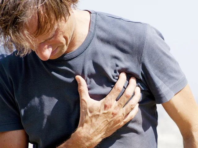
INTRODUCTION: Myocardial infarction (MI) is the formal term for what is commonly referred to as a heart attack. It normally results from a lack of blood flow (ischemia) and oxygen (hypoxia) to a region of the heart, resulting in death of the cardiac muscle cells. An MI often occurs when a coronary artery is blocked by the buildup of atherosclerotic plaque consisting of lipids, cholesterol and fatty acids, and white blood cells, primarily macrophages. It can also occur when a portion of an unstable atherosclerotic plaque travels through the coronary arterial system and lodges in one of the smaller vessels. The resulting blockage restricts the flow of blood and oxygen to the myocardium and causes death of the tissue.  MIs may be triggered by excessive exercise, in which the partially occluded artery is no longer able to pump sufficient quantities of blood, or severe stress, which may induce spasm of the smooth muscle in the walls of the vessel.

SIGNS AND SYMPTOMS: In the case of acute MI, there is often sudden pain beneath the sternum (retrosternal pain) called angina pectoris, often radiating down the left arm in males but not in female patients. Until this anomaly between the sexes was discovered, many female patients suffering MIs were misdiagnosed and sent home. In addition, patients typically present with difficulty breathing and shortness of breath (dyspnea), irregular heartbeat (palpations), nausea and vomiting, sweating (diaphoresis), anxiety, and fainting (syncope), although not all of these symptoms may be present. Many of the symptoms are shared with other medical conditions, including anxiety attacks and simple indigestion, so differential diagnosis is critical. It is estimated that between 22 and 64 percent of MIs present without any symptoms.

ASSESSMENT AND DIAGNOSIS: An MI can be confirmed by examining the patient’s ECG, which frequently reveals alterations in the ST and Q components. Some classification schemes of MI are referred to as ST-elevated MI (STEMI) and non-elevated MI (non-STEMI). In addition, echocardiography or cardiac magnetic resonance imaging may be employed.

Blood tests: Common blood tests indicating an MI include elevated levels of creatine kinase MB (an enzyme that catalyzes the conversion of creatine to phosphocreatine, consuming ATP) and cardiac troponin (the regulatory protein for muscle contraction), both of which are released by damaged cardiac muscle cells. 

MEDICAL MANAGEMENT: Immediate treatments for MI are essential and include administering supplemental oxygen, aspirin that helps to break up clots, and nitroglycerine administered sublingually (under the tongue) to facilitate its absorption. Despite its unquestioned success in treatments and use since the 1880s, the mechanism of nitroglycerine is still incompletely understood but is believed to involve the release of nitric oxide, a known vasodilator, and endothelium-derived releasing factor, which also relaxes the smooth muscle in the tunica media of coronary vessels.

Longer-term treatments include injections of thrombolytic agents such as streptokinase that dissolve the clot, the anticoagulant heparin, balloon angioplasty and stents to open blocked vessels, and bypass surgery to allow blood to pass around the site of blockage. If the damage is extensive, coronary replacement with a donor heart or coronary assist device, a sophisticated mechanical device that supplements the pumping activity of the heart, may be employed.

RISK FACTORS: Important risk factors for MI include cardiovascular disease, age, smoking, high blood levels of the low-density lipoprotein (LDL, often referred to as “bad” cholesterol), low levels of high-density lipoprotein (HDL, or “good” cholesterol), hypertension, diabetes mellitus, obesity, lack of physical exercise, chronic kidney disease, excessive alcohol consumption, and use of illegal drugs.


RELATED;

1. HYPERTENSION  2. DIABETES MELLITUS  3. OBESITY

REFERENCES

5 WONDERS OF THE HUMAN LIFE

WONDERS OF THE HUMAN LIFE: 
The human life is one of it's kind and it has several wonders that we hardly think about.  The human body is seen as wholly and a single entity that may seem to have no divisions but unlike bacteria, and other microbes, our bodies are complex and made up of organs and systems.  From head to toe, the human body is seen as a whole entity almost made up of no special components and functioning is far from reach to a naked eye.  From birth to adulthood, and including the very many processes going on in the human body, including in-utero, this article is going to be looking at the most incredible circumstances that every mankind should think about.  These are physiological processes, anatomical formations or just abnormal changes and deviations in the human body.

1.  THE CARDIOVASCULAR SYSTEM:
Our very first wonder is located in the cardiovascular system and specifically the heart.  The human heart is formed in the 4th week of the embryonic life in the uterus and by the fifth week, it has started pumping although there are no developed blood vessels and blood or chambers at a time.  This body's vital organ the size of your fist in an average adult individual, continues to pump unstoppably throughout life and it will only stop at the point of deathWith it's ability to pump blood through more than 75Kg of an average individual, this backbone of life can make the approximate 5.5-7 liters of blood for an average individual circulate the entire body in only about 75 seconds.

It is also astonishing to know that the human heart is capable of generating nerve impulses on it's own irrespective of the input from the central nervous system and can remain pumping for more than 9 minutes outside the human body as long as it is immersed in a solution containing glucose.  There is too much I have discussed about the human heart and to read more about the human heart, click here.

2.  THE PARALLEL CONNECTION OF BLOOD SUPPLY: 
It is astonishing to know that all organs of the human body supplied blood via a parallel connection.  This means that every organ receives it's own branch of blood vessel from the main blood vessel, the aorta.  There are only two known instances where a series connection is available and in that sense, blood from one organ will pass through another organ and we call the blood vessel in that case, a portal vein.  This is between the hypothalamus and pituitary in the first instance, and in that case we have the hypothalamic portal vein, then between the liver and the intestines, in which case we have the hepatic portal vein.  I have recently discussed about the hepatic circulation and you can read more about it here.

3.  THE CONTINUOUS DEMAND FOR INTAKE OF FOOD: 
The other most unnotably wonderful fact about the human body is the endless demand to eat.  Our bodies start the continuous input of food in our mother's wombs when we are feeding via the umbrical cord and it continues to breast milk feeding after birth linking the same body that natured us, and then food is introduced at advancing age and the eating habit continues.  This unstoppable and continuous process of the human body continues throughout life and it is the only way the body get nourished with nutrients.  It is therefore our role and responsibility to feed our bodies.

4.  THE MATERNAL-FETAL CIRCULATION: 
Our fourth wonder of the human body lies between the blood supply of a pregnant mother and her fetus.  It is known by very few that the maternal blood does not mix with that of the fetus during pregnancy and both the mother and the growing fetus both rely on independent circulatory systems.  It could be proved wrong or misunderstood but to clearly state the evidence, we look at the maternal blood group versus the fetal blood group which may be totally different and this would trigger death from blood incompatibilities if both blood systems were communicating directly.

During embryonic development, there develops an organ known as the placenta on the inner walls of the uterus and this is where exchange of material happens.  Now depending on the nature of the substance, some will diffuse from the mother to the fetal side and vice versa.

5.  THE GIT TRANSIT TIME: 
The human gastrointestinal tract is about 9 meters long from the mouth to the anal canal.  This system is the only one responsible for nourishing the body with ions and nutrient, following input and digestion of food.  The wonders of this multi-organ system of varying tissues and diameters is that food will stay in it for about 36 hours on average following ingestion.  But of course this time will depend on the type of food eaten and the extent of exercise carried out by the individual plus some other factors such as disease state.   It is this time that all the possible nutrients will be absorbed. The deviation from that, will be of two extents; either increased transit time in which food will overstay in it, or reduced transit time in which food will harry leaving the system.  The two extremes have two consequences as we can see; in case of increased time, the fecal matter will be compacted and become super solid leading to a condition known as constipation.  In case or reduced transit time, food will harry leaving the system and with it, goes the precious nutrients and water leading to a condition known as diarrhea.

RELATED;

July 31, 2025

VITAMINS AND MINERALS


INTRODUCTION: Vitamins are organic molecules needed in very small amounts for normal body functioning. Some vitamins are coenzymes; that is, they are necessary for the functioning of certain enzymes. Others are antioxidant vitamins, including vitamins C, E, and betacarotene, which is a precursor for the synthesis of vitamin A. Antioxidants prevent damage from free radicals, which are molecules that contain an unpaired electron and are highly reactive. The reactions of free radicals can damage DNA, cell membranes, and the cell organelles.

FORMATION OF FREE RADICALS: Free radicals are formed during some normal body reactions, but smoking and exposure to pollution will increase their formation. Antioxidant vitamins combine with free radicals before they can react with cellular components. Plant foods are good sources of these vitamins.

DEFICIENCY OF VITAMINS: Deficiencies of vitamins often result in diseases including but not limited to: vitamin C deficiency and scurvy, for example. Other deficiency diseases that have been known for decades include pellagra which is due to lack of niacin, beri-beri which is due to lack of riboflavin, pernicious anemia for lack of vitamin B12, and rickets for lack of vitamin D. More recently the importance of folic acid (folacin) for the development of the fetal central nervous system has been recognized. Adequate folic acid during pregnancy can significantly decrease the chance of spina bifida also known as, open spinal column and anencephaly which is the absence of the cerebrum and always fatal in a fetus. All women should be aware of the need for extra folic acid during pregnancy.

MINERALS IN THE HUMAN BODY: Minerals are simple inorganic chemicals and have a variety of functions, many of which you are already familiar with. Among some of the most important minerals we have Sodium and potassium which are very important in the normal functioning of the nervous system and the heart, Calcium important in bone formation, Magnesium and phosphorous as we shall be discussing them in details later.


RELATED;

1. VITAMIN A

2.  VITAMIN C

3.  MAGNESIUM

4.  DYNAMICS OF THE HUMAN BODY

REFERENCES

June 17, 2025

INTRAVENOUS INFUSION OF FLUIDS


INTRODUCTION: When fluid output exceeds fluid intake, volume deficits may result. Shock, dehydration, or electrolyte loss may occur; large deficits are fatal, unless treated. The following are some common reasons for fluid depletion: 

1) Loss of gastrointestinal (GI) fluids due to vomiting, diarrhea, chronic laxative use, or GI suctioning. 

2) Excessive sweating during hot weather, athletic activity, or prolonged fever. 

3) Severe burns. 

4) Hemorrhage. 

5) Excessive diuresis due to diuretic therapy or uncontrolled diabetic ketoacidosis.

PURPOSE OF ADMINISTERING IV FLUIDS: The immediate goal in treating a volume deficit disorder is to replace the depleted fluid. In non-acute circumstances, this may be achieved by drinking more liquids or by administering fluids via a feeding tube. In acute situations, IV fluid therapy is indicated. Regardless of the route, careful attention must be paid to restoring normal levels of blood elements and electrolytes as well as fluid volume. IV replacement fluids are of two basic types namely; crystalloids and colloids.

CRYSTALLOIDS: Crystalloids are IV solutions that contain electrolytes and other substances that closely mimic the body’s ECF. They are used to replace depleted fluids and to promote urine output. Crystalloid solutions are capable of quickly diffusing across membranes, leaving the plasma and entering the interstitial fluid and ICF. It is estimated that two thirds of infused crystalloids will distribute in the interstitial space.

COMPONENTS OF IV FLUIDS: Isotonic, hypotonic, and hypertonic solutions are available for that purpose. Sodium is the most common crystalloid added to solutions. Some crystalloids contain dextrose, a form of glucose, commonly in concentrations of 2.5%, 5%, or 10%. Dextrose is added to provide nutritional value: 1 L of 5% dextrose supplies 170 calories. In addition, water is formed during the metabolism of dextrose, enhancing the rehydration of the patient. When dextrose is infused, it is metabolized, and the solution becomes hypotonic.

EFFECTS OF IV FLUIDS: Infusion of crystalloids will increase total fluid volume in the body, but the compartment that is most expanded depends on the solute in this case, sodium, concentration of the fluid administered. Isotonic crystalloids can expand the circulating intravascular (plasma) fluid volume without causing major fluid shifts between compartments. Isotonic crystalloids such as normal saline are often used to treat fluid loss due to vomiting, diarrhea, or surgical procedures, especially when the blood pressure is low.

Because isotonic crystalloids can rapidly expand circulating blood volume, care must be taken not to cause fluid overload in the patient. Infusion of hypertonic crystalloids expands plasma volume by drawing water away from the cells and tissues.


RELATED;

1.  PLASMA VOLUME EXPANDERS

2.  BODY FLUIDS

3.  ANATOMY AND PHYSIOLOGY

REFERENCES

NICOTINE


INTRODUCTION:
  Nicotine is sometimes considered a CNS stimulant, and although it does increase alertness, its actions and long-term consequences place it in a class by itself. Nicotine is unique among abused substances in that it is legal, strongly addictive, and highly carcinogenic. Furthermore, use of tobacco can cause harmful effects to those in the immediate area who breathe secondhand smoke.  Patients often do not consider tobacco use as substance abuse. 

Tobacco Use and Nicotine: The most common method by which nicotine enters the body is through the inhalation of cigarette, pipe, or cigar smoke. Tobacco smoke contains more than 1,000 chemicals, a significant number of which are carcinogens. The primary addictive substance present in cigarette smoke is nicotine. Effects of inhaled nicotine may last from 30 minutes to several hours. Nicotine affects many body systems including the nervous, cardiovascular, and endocrine systems. 

Pharmacodynamics:  Nicotine stimulates the CNS directly, causing increased alertness and ability to focus, feelings of relaxation, and light-headedness. The cardiovascular effects of nicotine include an accelerated heart rate and increased blood pressure, caused by activation of nicotinic receptors located throughout the autonomic nervous system.

These cardiovascular effects can be particularly serious in patients taking oral contraceptives: The risk of a fatal heart attack is five times greater in smokers than in nonsmokers. Muscular tremors may occur with moderate doses of nicotine, and convulsions may result from very high doses. Nicotine affects the endocrine system by increasing the basal metabolic rate, leading to weight loss. Nicotine also reduces appetite. Chronic smoking leads to bronchitis, emphysema, and lung cancer. Both psychological and physical dependence occur relatively quickly with nicotine. Dependence

Once started on tobacco, patient tend to continue their drug use for many years, despite overwhelming medical evidence that the quality of life will be adversely affected and their life span shortened. Discontinuation results in agitation, weight gain, anxiety, headache, and an extreme craving for the drug.  Although nicotine replacement patches and gum assist patients in dealing with the unpleasant withdrawal symptoms, only 25% of patients who attempt to stop smoking remain tobacco-free a year later.

RELATED;
1.  CAFFEINE  

June 08, 2025

BREAST EXAMINATION


OBJECTIVES OF THE DISCUSSION:  By the end of this discussion, the reader/learner/medical student will be able to;
1.  List the importance of conducting breast examination
2.  Describe the normal findings that follows breast examination
3.  Explain the possible abnormal finding that can follow breast examination

INTRODUCTION: The breast examination by a physician remains the best means of early detection of breast cancer when combined with appropriately scheduled mammography. The results of the breast examination may be expressed by description or diagram, or both, usually with reference to the quadrants and tail region of the breast or by allusion to the breast as a clock face with the nipple at the center.

INSPECTION OF THE BREASTS: The breasts are first examined by inspection, with the patient’s arms at her sides, and then with her hands pressed against her hips, and/or with her arms raised over her head. If the patient’s breasts are especially large and pendulous, she may be asked to lean forward so that the breasts hang free of the chest, facilitating inspection.

FINDINGS: Tumors often distort the relations of these tissues, causing disruption of the shape, contour, and symmetry of the breast or position of the nipple. Some asymmetry of the breasts is common, but marked differences or recent changes deserve further evaluation.

1. Discolorations or ulcerations of the skin of the breast, areola, or nipple, or edema of the lymphatics that causes a leathery puckered appearance of the skin are abnormal.

2. A clear or milky breast discharge is usually bilateral and associated with stimulation or elevated prolactin levels also medically termed galactorrhea.

3. Bloody discharge from the breast is abnormal and usually unilateral; it usually does not represent carcinoma, but rather inflammation of a breast structure with intraductal papilloma is often found. Evaluation is necessary to exclude malignancy.

4. Pus usually indicates infection, although an underlying tumor may be encountered.

PALPATION OF THE BREASTS: Palpation follows inspection, first with the patient’s arms at her sides and then with the arms raised over her head. This part of the examination is usually done with the patient in the supine position. The patient may also be seated, with her arm resting on the examiner’s shoulder or over her head, for examination of the most lateral aspects of the axilla. Palpation should be done with slow, careful maneuvers, using the flat part of the fingers rather than the tips. The fingers are moved up and down in a wavelike motion, moving the tissues under them back and forth, so that any breast masses that are present can be more easily felt.

FINDINGS: If masses are found, their size, shape, consistency, and mobility as well as their position should be determined. Women with large breasts may have a firm ridge of tissue located transversely along the lower edge of the breast. This is the inframammary ridge and is a normal finding. The examination is concluded with gentle pressure inward and then upward at the sides of the areola to express fluid. If fluid is noted on inspection or is expressed, it should be sent for culture and sensitivity investigations.

RELATED;

1.  PELVIC INFLAMMATORY DISEASE

2.  OXYTOCIN

3.  OVULATION AND MENSTRUAL CYCLE

REFERENCES

April 13, 2025

LUNG CANCER


INTRODUCTION: Lung cancers arise from a single transformed epithelial cell in the tracheobronchial airways. A carcinogen such as; cigarette smoke, radon gas and other occupational and environmental agents, damages the cell, causing abnormal growth and development into a malignant tumor. Most lung cancers are classified into one of two major categories: small cell lung cancer which contribute 15% to 20% of tumors, and non–small cell lung cancer that contribute approximately 80% of tumors. Most small cell cancers arise in the major bronchi and spread by infiltration along the bronchial wall.

RISK FACTORS OF LUNG CANCER: Risk factors include tobacco smoke and especially second-hand also known as passive smoke, environmental and occupational exposures, gender, genetics, and dietary deficits. Other factors that have been associated with lung cancer include genetic predisposition and underlying respiratory diseases, such as chronic obstructive pulmonary disease (COPD) and tuberculosis (TB).

CLINICAL MANIFESTATIONS: Lung cancer often develops insidiously and is asymptomatic until late in its course. Signs and symptoms depend on location, tumor size, degree of obstruction, and existence of metastases to regional or distant sites.

1.  Most common symptom is cough or change in a chronic cough.

2.  Dyspnea may occur early in the disease.

3.  Hemoptysis or blood-tinged sputum may be expectorated.

4.  Chest pain or shoulder pain may indicate chest wall or pleural involvement. Pain is a late symptom and may be related to bone metastasis.

5.  Recurring fever may be an early symptom.

Chest pain, tightness, hoarseness, dysphagia, head and neck edema, and symptoms of pleural or pericardial infusion exist if the tumor spreads to adjacent structures and lymph nodes.  Common sites of metastases are lymph nodes, bone, brain, contralateral lung, adrenal glands, and liver.  Weakness, anorexia, and weight loss may appear.

ASSESSMENT AND DIAGNOSTIC METHODS: Chest x-ray, CT scans, bone scans, abdominal scans, PET scans, liver ultrasound, and MRI.  Sputum examinations, fiberoptic bronchoscopy, transthoracic fine-needle aspiration, endoscopy with esophageal ultrasound, mediastinoscopy or mediastinotomy, and biopsy.  Pulmonary function tests, ABG analysis scans, and exercise testing.  Staging of the tumor refers to the size of the tumor, its location, whether lymph nodes are involved, and whether the cancer has spread.

MEDICAL MANAGEMENT: The objective of management is to provide a cure if possible. Treatment depends on cell type, stage of the disease, and physiologic status. Treatment may involve surgery, radiation therapy, or chemotherapy—or a combination of these.

RELATED;

1.  THE ORIGIN OF CANCER

2.  GASEOUS EXCHANGE

3.  ACIDITY AND ALKALINITY OF BODY SYSTEMS

REFERENCES

MOST FREQUENTLY READ