INTRODUCTION:
The two kidneys are located in the upper abdominal cavity on
either side of the vertebral column, behind the peritoneum. The upper
portions of the kidneys rest on the lower surface of the diaphragm
and are enclosed and protected by the lower rib cage. The kidneys are
embedded in adipose tissue that acts as a cushion and is in turn
covered by a fibrous connective tissue membrane called the renal
fascia, which helps hold the kidneys in place.
BASIC
ANATOMY OF THE KIDNEY: Each kidney has an indentation called the
hilus on its medial side. At the hilus, the renal artery enters the
kidney, and the renal vein and ureter emerge. The renal artery is a
branch of the abdominal aorta, and the renal vein returns blood to
the inferior vena cava. The ureter carries urine from the kidney to
the urinary bladder.
INTERNAL
STRUCTURE OF THE KIDNEY: In a coronal or frontal section of the
kidney, three areas can be distinguished. The lateral and middle
areas are tissue layers, and the medial area at the hilus is a
cavity. The outer tissue layer is called the renal cortex; it is made
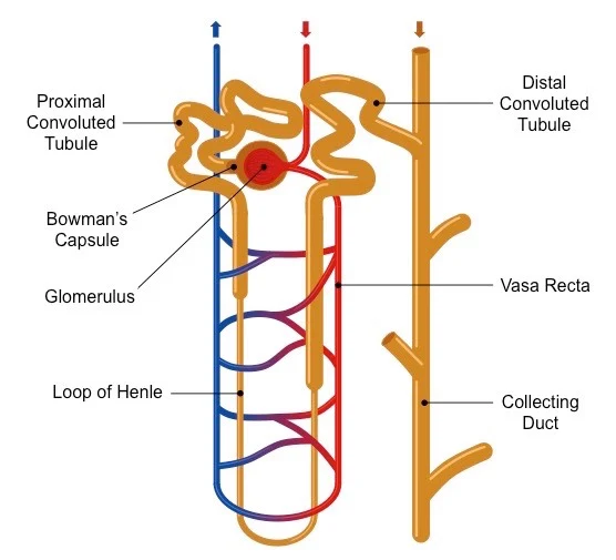
of renal corpuscles and convoluted tubules. These are parts of the
nephron and are described in details in our next discussion. The
inner tissue layer is the renal medulla, which is made of loops of
Henle and collecting tubules which are also, parts of the nephron.
The renal medulla consists of wedge-shaped pieces called renal
pyramids. The tip of each pyramid is its apex or papilla. The third
area is the renal pelvis, and this is not a layer of tissues, but
rather a cavity formed by the expansion of the ureter within the
kidney at the hilus. Funnel shaped extensions of the renal pelvis,
called calyces or in singular: calyx, enclose the papillae of the
renal pyramids. Urine flows from the renal pyramids into the calyces,
then to the renal pelvis and out into the ureter.
RELATED;
1. RENAL FAILURE
2. WATER INTAKE AND OUTPUT
3. ALDOSTERONE
4. ADRENAL GLANDS
4. ACIDITY AND ALKALINITY OF BODY SYSTEMS
REFERENCES


No comments:
Post a Comment