Wednesday, November 10, 2021

TISSUES


INTRODUCTION:
A tissue is a group of cells with similar structure and function. The tissue contributes to the functioning of the organs in which it is found. The four groups of tissues are epithelial, connective, muscle, and nerve tissue.
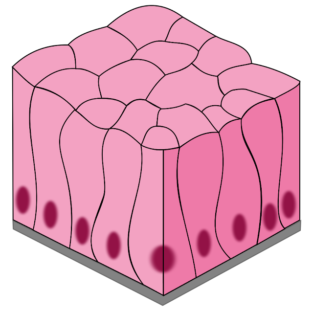
EPITHELIAL TISSUE: Epithelial tissues are found on surfaces as either coverings for the outer surfaces, or linings for the inner surfaces. Because they have no capillaries of their own, epithelial tissues receive oxygen and nutrients from the blood supply of the connective tissue beneath them. Many epithelial tissues are capable of secretion and may be called glandular epithelium, or more simply, glands.  Classification of the epithelial tissues is based on the type of cell of which the tissue is made, its characteristic shape, and the number of layers of cells. There are three distinctive shapes: squamous cells are flat, cuboidal cells are cube shaped, and columnar cells are tall and narrow. “Simple” is the term for a single layer of cells, and “stratified” means that many layers.

SIMPLE SQUAMOUS EPITHELIUM: Simple squamous epithelium is a single layer of flat cells. These cells are very thin and very smooth. The alveoli (air sacs) of the lungs are simple squamous epithelium. The thinness of the cells permits the diffusion of gases between the air and blood. Another location of this tissue is capillaries, the smallest blood vessels. Capillary walls are only one cell thick, which permits the exchange of gases, nutrients, and waste products between the blood and tissue fluid. The interior surface of capillaries is also very smooth. These cells continue as the lining of the arteries, veins, and heart; this is important because it prevents abnormal blood clotting within blood vessels.

STRATIFIED SQUAMOUS EPITHELIUM: Stratified squamous epithelium consists of many layers of mostly flat cells, although lower cells are rounded. Mitosis takes place in the lowest layer to continually produce new cells to replace those worn off the surface. This type of epithelium makes up the epidermis of the skin, where it is called keratinizing because the protein keratin is produced, and the surface cells are dead. Stratified squamous epithelium of the non-keratinizing type lines the oral cavity, the esophagus, and, in women, the vagina. In these locations the surface cells are living and make up the mucous membranes of these organs. In all of its body locations, this tissue is a barrier to microorganisms because the cells of which it is made are very close together.

TRANSITIONAL EPITHELIUM: Transitional epithelium is a type of stratified epithelium in which the surface cells change shape from round to squamous. The urinary bladder is lined with transitional epithelium. When the bladder is empty, the surface cells are rounded. As the bladder fills, these cells become flattened. Transitional epithelium enables the bladder to fill and stretch without tearing the lining.

SIMPLE CUBOIDAL EPITHELIUM: Simple cuboidal epithelium is a single layer of cube-shaped cells. This type of tissue makes up the functional units of the thyroid gland and salivary glands. These are examples of glandular epithelium; their function is secretion. In these glands the cuboidal cells are arranged in small spheres and secrete into the cavity formed by the sphere. In the thyroid gland, the cuboidal epithelium secretes the thyroid hormones; thyroxine is an example. In the salivary glands the cuboidal cells secrete saliva. Cuboidal epithelium also makes up portions of the kidney tubules. Here the cells have microvilli, and their function is the reabsorption of useful materials back to the blood.

SIMPLE COLUMNAR EPITHELIUM: Columnar cells are taller than they are wide and are specialized for secretion and absorption. The stomach lining is made of columnar epithelium that secretes gastric juice for digestion. The lining of the small intestine secretes digestive enzymes, but these cells also absorb the end products of digestion from the cavity of the intestine into the blood and lymph. To absorb efficiently, the columnar cells of the small intestine have microvilli, which you may recall are folds of the cell membrane on their free surfaces. These microscopic folds greatly increase the surface area for absorption. Yet another type of columnar cell is the goblet cell, which is a unicellular gland. Goblet cells secrete mucus and are found in the lining of the intestines and the lining of parts of the respiratory tract such as the trachea.

CILIATED EPITHELIUM: Ciliated epithelium consists of columnar cells that have cilia on their free surfaces. Ciliated epithelium lines the nasal cavities, larynx, trachea, and large bronchial tubes. The cilia sweep mucus, with trapped dust and bacteria from the inhaled air, toward the pharynx to be swallowed. Bacteria are then destroyed by the hydrochloric acid in the stomach. The air that reaches the lungs is almost entirely free of pathogens and particulate pollution. Another location of ciliated epithelium in women is the lining of the fallopian tubes. The cilia here sweep the ovum, which has no means of self-locomotion, toward the uterus.

RELATED;

1.  THE CELL MEMBRANE

2.  ANATOMY AND PHYSIOLOGY

REFERENCES

No comments:

Post a Comment

MOST FREQUENTLY READ