Showing posts with label August 2021. Show all posts

Sunday, August 22, 2021

PYRAZINAMIDE


INTRODUCTION:  Pyrazinamide (PZA) is a relative of nicotinamide. It is stable and slightly soluble in water. It is inactive at neutral pH, but at pH 5.5 it inhibits tubercle bacilli at concentrations of approximately 20 mcg/mL. The drug is taken up by macrophages and exerts its activity against mycobacteria residing within the acidic environment of lysosomes.

MECHANISM OF ACTION:  Pyrazinamide is converted to pyrazinoic acid, the active form of the drug by mycobacterial pyrazinamidase. The specific drug target is unknown, but pyrazinoic acid disrupts mycobacterial cell membrane metabolism and transport functions.

RESISTANCE TO THE DRUG:  Resistance may be due to impaired uptake of pyrazinamide or mutations in pncA that impair conversion of pyrazinamide to its active form.

PHARMACOKINETICS:  Serum concentrations of 30–50 mcg/mL at 1–2 hours after oral administration are achieved with dosages of 25 mg/kg/d. Pyrazinamide is well absorbed from the gastrointestinal tract and widely distributed in body tissues, including inflamed meninges. The half-life is 8–11 hours. The parent compound is metabolized by the liver, but metabolites are renally cleared; therefore, pyrazinamide should be administered at 25–35 mg/kg three times weekly (not daily) in hemodialysis patients and those in whom the creatinine clearance is less than 30 mL/min.  In patients with normal renal function, a dose of 40–50 mg/kg is used for thrice weekly or twice-weekly treatment regimens.

CLINICAL USES:  Pyrazinamide is an important front-line drug used in conjunction with isoniazid and rifampin in short-course of 6-month regimens as a “sterilizing” agent active against residual intracellular organisms that may cause relapse. Tubercle bacilli develop resistance to pyrazinamide fairly readily, but there is no cross-resistance with isoniazid or other antimycobacterial drugs.

ADVERSE REACTIONS:  Major adverse effects of pyrazinamide include hepatotoxicity (in 1–5% of patients), nausea, vomiting, drug fever, and hyperuricemia. The latter occurs uniformly and is not a reason to halt therapy. Hyperuricemia may provoke acute gouty arthritis.


RELATED;

1.  TUBERCULOSIS

2. ISONIAZID

3. ETHAMBUTOL

4. RIFAMPIN

5. GOUT

6. MEDICAL CONDITIONS

REFERENCES

Saturday, August 21, 2021

NOTES AND TEXTBOOKS

NOTES AND TEXTBOOKS:  From this page, download notes and textbooks in form of PDFs, PowerPoint presentation and word document.

1.  Obstetrics and gynecolog

a)  First aid for the obstetrics and gynecology clerkship

Download pdf

b)  Blueprints obstetrics and gynecology

Download pdf


2.  Microbiology

a)  Ananthanarayan and Paniker’s Textbook of Microbiology

Download pdf


3.  Biochemistry

a)  Integrative human biochemistry


4.  Medical research

i)  Qualitative research ppt

ii)  Research Methodology a step by step

Download pdf

iii)  research proposal template

Download pdf

iv)  Reasearch methodology 

Download powerpoint


5.  Community Health

i)  Introduction to community Health


6.  Biostatistics and demography

i)  An overview of biostatistics

ii)  Biostatistics and demography for medical students


7.  


8.  Clinical skills and practice

a) Textbook of clinical skills

Download pdf


9.  Pharmacology

i)  Basic and clinical pharmacology 24th edition

ii)  Pharmacology for nurses by M. Adams  4th edition

iii)  Stages of drug discovery

Sunday, August 15, 2021

WOUND HEALING BY SECONDARY INTENTION


INTRODUCTION:  Wound Healing by Second Intention.  This is also known as Secondary Union.  This type of healing occurs when injuries result in more extensive loss of tissues, such as with infarction, inflammatory ulceration, and large surface wounds. In these situations, due to the large tissue defects, repair by regeneration is minimal and the defect is filled with granulation tissue.  
Second intention healing differ from first intention healing in several ways as described below;  First the greater injury induce a more intense inflammatory response. Secondly, much more granulation tissue is formed, and thirdly wounds that are repaired by second intention healing undergo a phenomenon known as "wound contraction" whereby specialized granulation tissue fibroblasts called myofibroblasts contract and dramatically reduce the size of the wound.

RELATED;

Saturday, August 14, 2021

CHOLINOMIMETICS

Introduction: The actions of acetylcholine released from autonomic and somatic motor nerves are terminated by enzymatic hydrolysis of the molecule. Hydrolysis is accomplished by the action of the enzyme acetylcholinesterase, which is present in high concentrations in cholinergic synapses. The indirect-acting cholinomimetics have their primary effect at the active site of this enzyme, although some also have direct actions at nicotinic receptors. Receptors in the Central nervous system

Structure: There are three chemical groups of cholinesterase inhibitors: (1) simple alcohols bearing a quaternary ammonium group, such as, edrophonium; (2) carbamic acid esters of alcohols having quaternary or tertiary ammonium groups (carbamates, such as, neostigmine); and (3) organic derivatives of phosphoric acid (organophosphates, such as, echothiophate). Toxicity due toorganophosphates

Pharmacodynamics: Mechanism of Action: Acetylcholinesterase is the primary target of these drugs, but butyrylcholinesterase is also inhibited. Acetylcholinesterase is an extremely active enzyme. In the initial catalytic step, acetylcholine binds to the enzyme’s active site and is hydrolyzed, yielding free choline and the acetylated enzyme. In the second step, the covalent acetyl-enzyme bond is split, with the addition of water (hydration). All the cholinesterase inhibitors increase the concentration of endogenous acetylcholine at cholinoceptors by inhibiting acetylcholinesterase. The organophosphate inhibitors are sometimes referred to as “irreversible” cholinesterase inhibitors, and edrophonium and the carbamates are considered “reversible” inhibitors because of the marked differences in duration of action. However, the molecular mechanisms of action of the three groups do not support this simplistic description.

RELATED;

1.  THE NERVE IMPULSE PROPAGATION

REFERENCES

HUMAN LEUKOCYTE ANTIGENS (HLA)


INTRODUCTION:  Human leukocyte antigens (HLA) are antigens on White Blood Cells that are representative of the antigens present on all the cells of an individual.  Leukocytes  These are our “self” antigens that identify cells that belong in the body. Recall that in the ABO blood group of Red Blood Cells, there are only two antigens, A and B, and four possible types: A, B, AB, and O. The ABO blood grouping:  Red blood cells:  HLA antigens are also given letter names. HLA A, B, and C are called class I proteins, with from 100 to more than 400 possibilities for the specific protein each can be. The several class II proteins are given various D designations and, again, there are many possibilities for each. Each person has two genes for each HLA type, because these types are inherited, just as RBC types are inherited.

DIFFERENCES IN HLA:  Members of the same family may have some of the same HLA types, and identical twins have exactly the same HLA types. The purpose of the HLA types is to provide a “self” comparison for the immune system to use when pathogens enter the body. The T lymphocytes compare the “self” antigens on macrophages to the antigens on bacteria and viruses. Because these antigens do not match ours, they are recognized as foreign; this is the first step in the destruction of a pathogen.

HLA AND ORGAN TRANSPLANTS:  The surgical transplantation of organs has also focused on the HLA. The most serious problem for the recipient of a transplanted heart or kidney is rejection of the organ and its destruction by the immune system. You may be familiar with the term tissue typing. This process involves determining the HLA types of a donated organ to see if one or several will match the HLA types of the potential recipient. If even one HLA type matches, the chance of rejection is lessened. Although all transplant recipients (except corneal) must receive immunosuppressive medications to prevent rejection, such medications make them more susceptible to infection.  The closer the HLA match of the donated organ, the lower the dosage of such medications, and the less chance of serious infections.  

HLA AND AUTOIMMUNITY:  Sometimes a virus enters the body and stimulates the immune system to produce antibodies. The virus is destroyed, but one of the person’s own antigens is so similar to the viral antigen that the immune system continues its activity and begins to destroy this similar part of the body.  This is a scenario known as autoimmunity.  Autoimmunity  Another possibility is that a virus damages a self-antigen to the extent that it is now so different that it will be perceived as foreign. These are two theories of how autoimmune diseases are triggered, which is the focus of much research in the field of immunology.  

RELATED;

1.  AUTOIMMUNE DISEASES

2. MAJOR HISTOCOMPATIBILITY COMPLEX

3. REFERENCES

Saturday, August 07, 2021

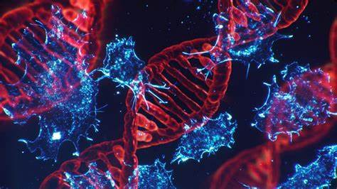
DNA, THE GENETIC MATERIAL


Introduction:  Imagine life without inheritance where everything can look like anything.  It would be so boring to have siblings resembling the neighbor and or resembling nothing, yet even traces of our lineages of succession would hardly be determined.  But thanks to nature, from the past until now, we can produce kids looking like the way our parents and grandfathers used to look like.  The matter is that we are not trying to be careful, but instead there is a road made for us:  The genetic predisposition, and the passing of genes from the cellular DNA.  Whether a woman or a man, strong or weak, lazy or powerful, there is a trail through which information is being passed in order to keep history.  In our discussion here, we are going to have a closer look at the genetic material also known as DNA and it's structural makeup.

STRUCTURE OF DNA:  Deoxyribonucleic acid (DNA) is composed of four deoxyribonucleotides, that is to say, deoxyadenylate (A), deoxyguanylate (G), deoxycytidylate (C), and thymidylate (T).  These units are combined through 3' to 5' phosphodiester bonds to polymerise into a long chain.  The nucleotide is formed by a combination of base + sugar + phosphoric acid.  The 3'-hydroxyl of one sugar is combined to the 5'-hydroxyl of another sugar through a phosphate group.  In this particular example, the thymidine is attached to cytidine and then cytidine to adenosine through phospho-diester linkages.  In the DNA, the base sequence is of paramount importance. The genetic information is coded in the specific sequence of bases; if the base is altered, the information is also altered.  The deoxyribose and phosphodiester linkages are the same in all the repeating nucleotides.  [RNA STRUCTURE AND FUNCTION]
Polarity of DNA molecule:  In the case of DNA, the base sequence is always written from the 5' end to the 3' end.  This is called the polarity of the DNA chain.
Watson-Crick Model of DNA Structure:  The salient features of Watson - Crick Model of DNA are given below:
Right handed double helix:  DNA consists of two polydeoxyribonucleotide chains twisted around one another in a right handed double helix similar to a spiral stair case.  The sugar and phosphate groups comprise the handrail and the bases jutting inside represent the steps of the staircase.  The bases are located perpendicular to the helix axis, whereas the sugars are nearly at right angles to the axis.

The base pairing rule:  Always the two strands are complementary to each other.  So, the adenine of one strand will pair with thymine of the opposite strand, while guanine will pair with cytosine.  The base pairing (A with T; G with C) is called Chargaff's rule, which states that the number of purines is equal to the number of pyrimidines.

Hydrogen bonding:  The DNA strands are held together mainly by hydrogen bonds between the purine and pyrimidine bases.  There are two hydrogen bonds between A and T while there are three hydrogen bonds between C and G.  The GC bond is therefore stronger than the AT bond.
Antiparallel: The two strands in a DNA molecule run antiparallel, which means that one strand runs in the 5' to 3' direction, while the other is in the 3' to 5' direction.  This is similar to a road divided into two, each half carrying traffic in the opposite direction.

RELATED;
1.  RNA

Friday, August 06, 2021

CARBON AS ACHEMICAL OF LIFE

CARBON AS ACHEMICAL OF LIFE:  Carbon is one of the most abundant atom on the planet available in both plants and animals.  There are very few if any substance of medical importance without at least a single atom of carbon in their chemical structure.  carbon is so common that we shall be meeting in almost all chemicals of life that we shall be looking at, and those we have already looked at.  It surfaces in all carbohydratesHydrocarbons, Lipids and even in gases such as Carbon dioxide and Carbon mono oxide.

BONDING OF CARBON: Carbon is capable of bonding with several other atoms in nature including but not limited to; Oxygen in which case it forms either Carbon dioxide or Carbon monoxide, Hydrogen in which case there is formation of hydrocarbons, Nitrogen where the is formation of cyanides among others.

The bonding of carbon is not limited to individual atoms but also, to functional groups and several compounds including the nitro group, carbon to carbon bonds and ions such as chloride (Cl-), Bromide (Br-) among others.

THE CARBON-CARBON BONDS........

RELATED;

MOST FREQUENTLY READ