Showing posts with label December 2022. Show all posts

Thursday, December 15, 2022

INFERTILITY


Definition: Infertility is defined as the absence of conception after at least 1 year of regular sexual intercourse.

Causes: Males are found to be solely responsible for 20-30% of infertility cases and these are related to issues such as, inadequate sperm count which contribute to 50% of cases.  For female infertility, about 40% of cases are due to ovulatory failure, about 40% are due to endometrial or tubal disease, about 10% are due to rarer causes such as, thyroid disease or hyperprolactinemia, and about 10% remain unexplained after full workup.

Pathophysiology of female infertility

Ovulatory Causes:  Infertility due to ovarian dysfunction can result from disorders of the hypothalamus or pituitary, resulting in inadequate gonadotropic stimulation of the ovary.  This can bring problems ranging from ovarian disorders, resulting either in inadequate secretory products or failure to ovulate; and occasionally from both types of disorder occurring at the same time. Correction of the underlying cause will often restore fertility. In many cases, the administration of exogenous gonadotropins will stimulate the ovaries to produce follicular growth. The oocytes can then be released in vivo and fertilized by intercourse or by artificial insemination.

Tubal and Pelvic Causes:  With normal follicles and reproductive neuroendocrine axis function, the major cause of infertility is an abnormality in the endometrium or fallopian tubes. Prior or ongoing pelvic infections, with adhesions or inflammation, can result in a failure of sperm or egg transport, a failure of implantation, or implantation in an inappropriate location (ectopic pregnancy).


RELATED;

1.  PELVIC INFLAMMATORY DISEASE

2.  OBSTETRICS AND GYNECOLOGY

3.  CONTRACEPTION

4.  MEDICAL CONDITIONS

REFERENCES


Wednesday, December 14, 2022

ATROPINE


Therapeutic Class: Antidote for anticholinesterase poisoning

Pharmacologic Class: Muscarinic cholinergic receptor blocker.

ACTIONS AND USES:  By occupying muscarinic receptors, atropine blocks the parasympathetic actions of Ach and induces symptoms of the fight-or-flight response. Most prominent are increased heart rate, bronchodilation, decreased motility in the GI tract, mydriasis, and decreased secretions from glands.  

At therapeutic doses, atropine has no effect on nicotinic receptors in ganglia or on skeletal muscle. Although atropine has been used for centuries for a variety of purposes, its use has declined in recent decades because of the development of safer and more effective medications. Atropine may be used to treat hypermotility diseases of the GI tract such as irritable bowel syndrome, to suppress secretions during surgical procedures, to increase the heart rate in patients with bradycardia, and to dilate the pupil during eye examinations. Once widely used to cause bronchodilation in patients with asthma, atropine is now rarely prescribed for this disorder. Atropine therapy is useful for the treatment of reflexive bradycardia in infants and infantile hypertrophic pyloric stenosis (IHPS).


ADMINISTRATION ALERTS

1.  Oral and subcutaneous doses are not interchangeable.

2. Monitor blood pressure, pulse, and respirations before administration and for at least 1 hour after subcutaneous administration.

3. Pregnancy category C.


ADVERSE EFFECTS:  The side effects of atropine limit its therapeutic usefulness and are predictable extensions of its autonomic actions. Expected side effects include dry mouth, constipation, urinary retention, and an increased heart rate. Initial CNS excitement may progress to delirium and even coma.

CONTRAINDICATIONS: Atropine is contraindicated in patients with glaucoma, because the drug may increase pressure within the eye. Atropine should not be administered to patients with obstructive disorders of the GI tract, paralytic ileus, bladder neck obstruction, benign prostatic hyperplasia, myasthenia gravis, cardiac insufficiency, or acute hemorrhage.

INTERACTIONS:  Drug–Drug: Drug interactions with atropine include an increased effect with antihistamines, TCAs, quinidine, and procainamide. Atropine decreases effects of levodopa.

TREATMENT OF OVERDOSE:  Overdose may cause CNS stimulation or depression. A short-acting barbiturate or diazepam (Valium) may be administered to control convulsions. Physostigmine is an antidote for atropine poisoning that quickly reverses the coma caused by large doses of atropine.


RELATED;

1.  ACETYLCHOLINE

2.  DIAZEPAM

3.  PHARMACOLOGY AND THERAPEUTICS

REFERENCES


Monday, December 12, 2022

THE RESEARCH STUDY SETTINGS


OBJECTIVES OF THE DISCUSSION:  By the end of this article, the reader/medical student will be able to;
1.  Outline the importance of a research study setting
2.  Explain the different types of research study setting

INTRODUCTION:  The setting is the location where a study is conducted. There are three common settings for conducting medical research: natural, partially controlled, and highly controlled. A natural setting, or field setting, is an uncontrolled, real-life situation or environment. Conducting a study in a natural setting means that the researcher does not manipulate or change the environment for the study. Descriptive and correlational quantitative studies, qualitative, mixed methods, and outcomes studies often are conducted in natural settings.

THE PARTIALLY CONTROLLED SETTING:  A partially controlled setting is an environment that the researcher manipulates or modifies in some way while conducting a study. This may include studies conducted in hospital settings.

THE HIGHLY CONTROLLED SETTING:  A highly controlled setting is a structured environment that often is artificially developed for the purpose of conducting research. Laboratories, research or experimental centers, and test or highly structured units in hospitals or other healthcare agencies are highly controlled settings. Often experimental and sometimes quasi-experimental studies are conducted in these types of settings. A highly controlled setting reduces the influence of extraneous variables, which enables researchers to examine accurately the effect of an intervention on an outcome.


RELATED;

1.  HOW TO WRITE A RESEARCH PROPOSAL

2.  COMPONENTS OF A RESEARCH PROPOSAL

3.  CHAPTER THREE:  RESEARCH METHODOLOGY

4.  STUDY DESIGNS

5.  MOST PREVAILING RESEARCH PROBLEMS 2022/2023

Saturday, December 10, 2022

CORTISOL


OBJECTIVES OF THE DISCUSSION:  By the end of this discussion, the reader/medical student will be able to;
1.  Describe cortisol and it's role in generation of stress situations

INTRODUCTION: In our discussion here, we will use cortisol as a representative of the group of hormones called glucocorticoids, because it is responsible for most of the actions of this group. Cortisol increases the use of fats and excess amino acids, in a metabolic pathway known as, gluconeogenesis for energy and decreases the use of glucose. This is called the glucose-sparing effect, and it is important because it conserves glucose for use by the brain.


SECRETION OF CORTISOL: Cortisol is secreted in any type of physiological stress situation including but no limited to; disease, physical injury, hemorrhage, fear or anger, exercise, and hunger. Although most body cells easily use fatty acids and excess amino acids in cell respiration, brain cells do not, so they must have glucose. By enabling other cells to use the alternative energy sources, cortisol ensures that whatever glucose is present will be available to the brain. 

Cortisol also has an anti-inflammatory effect. During inflammation, histamine from damaged tissues makes capillaries more permeable, and the lysosomes of damaged cells release their enzymes, which help break down damaged tissue but may also cause destruction of nearby healthy tissue. Cortisol blocks the effects of histamine and stabilizes lysosomal membranes, preventing excessive tissue destruction. Inflammation is a beneficial process up to a point, and is an essential first step if tissue repair is to take place. It may, however, become a vicious cycle of damage, inflammation, more damage, more inflammation, and so on in a positive feedback mechanism. Normal cortisol secretion seems to be the brake, to limit the inflammation process to what is useful for tissue repair, and to prevent excessive tissue destruction.


ADVERSE EFFECTS OF CORTISOL: Too much cortisol decreases the immune response, leaving the body susceptible to infection and significantly slowing the healing of damaged tissue. The direct stimulus for cortisol secretion is ACTH from the anterior pituitary gland, which in turn is stimulated by corticotropin releasing hormone (CRH) from the hypothalamus. CRH is produced in the physiological stress situations mentioned earlier. Although we often think of epinephrine as a hormone important in stress, cortisol is also important.


RELATED;

1. GLUCONEOGENESIS

2.  CATECHOLAMINES

3.  METABOLISM AND METABOLIC DISORDERS

4.  GLUCONEOGENESIS

5.  BIOCHEMISTRY

REFERENCES


Thursday, December 08, 2022

THE ANDROID DEBUG/DAEMON BRIDGE


INTRODUCTION: I am not a software developer, IT consult or a computer scientist but just an android enthusiast, who have used adb and fastboot commands for more than 3 years now and I acknowledge their usefulness. For the period I have been using smartphones and computers I have found out that when it comes to transferring files to and from phone, sometimes the conventional methods that were very effective in ordinarily button-based phones may not apply and may need sophisticated procedures with smartphones. But these are files that maybe needed urgently and you received them over WhatsApp, your email, or you just needed to share them with your worker-mate. I remember that in our ordinarily phones it was very easy once you connected the phone to your computer with a USB cable, then automatically the request would come “Use as a media device” or “Connect U-disk” and then the other would be “Connect USB for charging”. Now with smartphones things changed somewhere somehow and maybe we have not kept the norm of reading Google articles and docs plus relevant information about the specific phones from the manufacturers.

THE ADB COMMANDS: These ADB commands and ADB Shell commands work on all Android devices regardless of the device model or manufacturer. They can be used by people who are used to rooting their Android devices or customization. These are system utilities that provide access to the backend of Android when it is connected to a PC. These are also one of the constants in the Android ecosystem. Same commands will produce the same results regardless of the device you have, in most cases. ADB or Android Debug Bridge can be used when the Android device is turned on while Fastboot can be used while the device is in the Bootloader or Fastboot mode.  Today we’ll take a look at some useful ADB and Fastboot commands. Before you can make use of ADB and Fastboot commands, you’ll have to enable USB Debugging which is located under development options on your Android device. The option is found in the Developer options but if you don’t know how to get there, you can follow the link below because by default, the developer options are always hidden.  How to enable USB debugging in android

Besides, you’ll also need the ADB and Fastboot drivers on your Windows, Mac, or Linux computer. ADB commands are used when the device is powered on and you have access to the device. That is, you can unlock the device and grant ADB debugging permission to the computer. Listed below are some of the most common and useful ADB commands that you may need at some point.

1. adb devices: Shows all the devices connected to ADB. This command can be used to make sure your device is connected properly before performing any other operations. This command will display the list of all Android devices attached to your PC.  After entering the command and pressing "enter", an alphanumeric figure will be displayed in front of the word device.

2. adb connect ip_address_of_device: Using the ‘adb connect’ command, you can connect the IP address of your Android device to your computer.

3. adb reboot: ADB can be used to reboot or restart your device, useful when your hardware buttons aren’t working or if you’re already using ADB. You can also reboot directly into the recovery or bootloader using this command.


4. adb reboot bootloader: Reboots the device into the Fastboot or Bootloader mode.  Depending on the type of smartphone you are having, you will see a sign that you are in Fastboot mode after entering this command, and then start using the fastboot commands.


5. adb install: It can be used to install an APK file on your Android device from your computer. It is more convenient than the alternative, which is to transfer the APK to the device and then install it. This command is generally used by developers or debuggers.


6. i)  adb uninstall: Use the following command if you want to uninstall an app package.  But sometimes it may not be able to uninstall system apps in which case you will use the following command;

ii)  adb shell pm uninstall -k –-user 0

It should be noted that, following these last three commands you have put the package name.


7. adb sideload: Just as you can flash zip packages from a custom recovery, you can also do it via adb sideload, provided the recovery supports it. This way you can flash packages directly from your computer without having to transfer them first to your device or any external storage.

8. adb shell: The ‘adb shell‘ command starts the remote shell command console in the device and lets you control the device through it.

i) adb shell pm uninstall -k --user 0: You can uninstall or remove any system app installed on your Android device. This is the easiest way to uninstall bloatware. Just use the following command followed by the app package name.


9.  adb backup -apk -shared -all -f backup.ab:  This command can be used to backup the entire phone.


RELATED LINKS;

1.  Full use of adb commands

2.  Android to PC connectivity

3.  Software download links

4.  Gospel music downloads

Sunday, December 04, 2022

CO-ENZYMES


INTRODUCTION: Enzymes may be simple proteins, or complex enzymes, containing a non-protein part, called the prosthetic group. The prosthetic group is called the co-enzyme and therefore may be non-proteins in nature. Usually the prosthetic group is heat stable than the protein component of the enzyme. The protein part of the enzyme is then named the apo-enzyme which is heat labile and then the two portions combined together is called the holo-enzyme.

CLASSIFICATION OF CO-ENZYMES: Co-enzymes may be divided into two groups;

1) Those taking part in reactions catalyzed by oxidoreductases by donating or accepting hydrogen atoms or electrons.

2) Those co-enzymes taking part in reactions transferring groups other than hydrogen.

In the first group, the change occurring in the substrate is counter-balanced by the co-enzymes. Therefore, such co-enzymes may be considered as co-substrates or secondary substrates.


NICOTINAMIDE ADENINE DINUCLEOTIDE (NAD+): This is a co-enzyme synthesized from Nicotinamide, a member of vitamin B complex. The reversible reaction of lactate to pyruvate is catalyzed by the enzyme lactate dehydrogenase, but the actual transfer of hydrogen is taking place on the co-enzyme, NAD+.

In this case, two hydrogen atoms are removed from lactate, out of which one hydrogen and two electrons are accepted by the NAD+ to form NADH, and the remaining H+ is released into the surrounding medium.


SECOND GROUP OF CO-ENZYMES: These co-enzymes take part in reactions transferring groups other than hydrogen. A particular group or radical is transferred from the substrate to another substrate. Most of them belong to vitamin B complex group.


ADENOSINE TRIPHOSPHATE (ATP): ATP is considered to be the energy currency in the body. In the ATP molecule, the second and third phosphate bonds are ‘high energy' bonds. During the oxidation of food stuffs, energy is released, a part of which is stored as chemical energy in the form of ATP. The endergonic reactions are carried out with the help of energy released from hydrolysis of ATP.

METALLO-ENZYMES: These are enzymes which require certain metal ions for their activity. In certain cases, such as copper in Tyrosinase, the metal is tightly bound with the enzyme. In other cases, even without the metal ion, enzyme may be active; but when the metal ion is added, the activity is enhanced. They are called ion-activated enzymes, for example, calcium ions will activate pancreatic lipase.

Co-factors: The term co-factor is used as a collective term to include co-enzymes and metal ions. Co-enzyme is an organic co-factor.


RELATED;

1. LACTATE

2. PYRUVATE

3. BIOCHEMISTRY OF ENZYMES

4. BIOMOLECULES AND CHEMICALS OF LIFE

REFERENCES

Saturday, December 03, 2022

PLASMA PROTEINS


INTRODUCTION: Total blood volume is about 4.5 to 5 liters in adult human being. If blood is mixed with an anticoagulant and centrifuged, the cell components which iclude Red blood cells (RBC) and white blood cells (WBC) are precipitated. The supernatant is called plasma. About 55-60% of blood is made up of plasma. If blood is withdrawn without anticoagulant and allowed to clot, after about 2 hours liquid portion is separated from the clot. This defibrinated plasma is called serum, which lacks coagulation factors including prothrombin and fibrinogen.


PROTEINS IN BLOOD: Total protein content of normal plasma is 6 to 8 g/100 ml. The plasma proteins consist of albumin, which is about 3.5 to 5 g/dl, globulins which make up 2.5-3.5 g/dl and fibrinogen making it to about 200-400 mg/dl. Almost all plasma proteins, except immunoglobulins are synthesized in liver. Plasma proteins are generally synthesised on membrane-bound polyribosomes. Most plasma proteins are glycoproteins.


RELATED;

1. BLOOD AND IT’S COMPONENTS

2. PROTEINS

3. IMMUNOGLOBULINS

REFERENCES


Thursday, December 01, 2022

EDEMA


Objectives of the discussion:  By the end of this discussion, the learner/medical student will be able to;
1.  Explain the cause of the swelling of the different human body parts
2.  Describe the difference between systemic and localised edema

Introduction: Edema is an abnormal increase in the amount of tissue fluid, which may be localized or systemic. Sometimes edema is inapparent, and sometimes it is apparent as swelling. 

Localized edema follows injury and inflammation of a body part.  I have discussed a lot about inflammation and the drugs used to treat it.  You can read more about inflammation from the link in the related below.

Pathophysiology of edema: Spraining an ankle, for example, damages tissues that then release histamine. Histamine increases the permeability of capillaries, and more tissue fluid is formed. As tissue fluid accumulates, the ankle may become swollen.

Systemic edema:  Systemic edema is the result of an imbalance between the movement of water out of and into capillaries, that is, between filtration and osmosis. Excessive filtration will occur when capillary pressure rises. This may be caused by venous obstruction due to blood clots or by congestive heart failure.  Edema of this type is often apparent in the lower extremities. Systemic bacterial infections may increase capillary permeability, and loss of plasma to tissue spaces is one aspect of septicemia. In this situation, however, the edema is of secondary importance to the hypotension, which may be life-threatening. 

Insufficient osmosis, the return of tissue fluid into capillaries, is a consequence of a decrease in plasma proteins, especially albumin. This may occur in severe liver diseases such as cirrhosis, kidney disease involving loss of protein in urine, malnutrition, or severe burn injuries.  Because edema is a symptom rather than a disease, treatment is aimed at correcting the specific cause. If that is not possible, the volume of tissue fluid may be diminished by a low-salt diet and the use of diuretics


RELATED;

1. THE INFLAMMATORY PROCESS

2. BIOCHEMISTRY OF HISTAMINE

3. CONGESTIVE HEART FAILURE

4.  MEDICAL CONDITIONS

REFERENCES

Wednesday, December 09, 2020

FULL LIST OF ADB COMMANDS


ADB AND FASTBOOT COMMANDS:  The Android Debug or Daemon Bridge (ADB) is a very useful way of controlling your smartphone if you are android enthusiast.  When smartphones came, most of us just had to enjoy the slippery touch screen and the disappearance of many buttons.  It later then became evident that there were many developments that were not explained to non developers.  From controlling a few installed apps to unlocking bootloader and flashing custom ROMs and flushable zips, I have enjoyed using the ADB.  I a m not a developer or a tech supporter and I must warn you that as you use this piece of idea, Mega Mover Empire got all about it from Google.  Therefore you should use it with caution an make sure that you read the trusted sources of whatever you want to do with it and also, Google the exact specifications of your phone's model and manufacturer before doing anything.

Before you start using the adb commands, make sure USB debugging is enabled on your phone, you have the original USB cable on your phone, and you have installed Minimal adb and fastboot software on your computer.  Other sources recommend installing and using SDK platform tools however because I am running some of the last versions of Windows XP, somewhere somehow, I failed to run the platform tools on my gadget.  Either way you do it doesn't matter as long as you get everything right.  You can follow the link below to download Minimal adb and fastboot setup, and learn how to activate USB debugging.
What next? Run the installer and make sure you fully install Minimal adb and fastboot setup you have just downloaded. Right click on minimal adb and fastboot shortcut on your desktop and choose the option "run as administrator", then connect you smartphone to your PC using your original USB cable.  The minimal adb and fastboot setup tool lools pretty like the windows CMD commandline interface.  Then follow the description below.

ADB COMMANDS: 
Enter these commands as you see the response either from your devices.
1.  Adb devices:  This command can be used to check availability of the device.  After entering the command, press enter and look at the phone screen and PC screen.  If it is the first time for your smartphone to be connected for ADB on that PC, a popup message will appear on your Phone's screen prompting you to accept authorization.  Please do so, and then, an alphanumeric figure will appear on your PC screen behind the word device.  Then you know your phone is detected.

2.  Adb push:  This command can be used to send files from the PC to the smartphone.  Basically the way it does, you have to indicate the source of the file you are sending to the phone, and then the directory where you want it to go.  For example:  I am sending a folder named "DOWNLOADED" from my PC to my android SDCARD and the command will be;

adb push "C:\Users\Muwanga Godfrey\Desktop\DOWNLOADED" /sdcard

You can also click on the link below to see a video demonstration about the above command.

Transfer files from PC to phone using adb commands

3.  Adb pull:  This command can be used to transfer files from the smartphone to PC.  All you have to do, is to specify on the source of the file on the android phone and then, the destination at the PC.  For example:  I am copying photos taken by my android camera to my PC in a folder named "DOWNLOADED" on the desktop, and the command will be;

adb pull /sdcard/DCIM/Camera  "C:\Users\Muwanga Godfrey\Desktop\DOWNLOADED"

To view our YouTube video about this adb command, follow the link below.

How to transfer files from android to PC using adb commands

4.  Adb install:  With this command, you can install android apps on your smartphone.  This is especially important when the app you want to install does not require using data or internet, and you already have it on your PC.  Again you have to specify on the location of the app on the PC, and end with the extension ".apk" after the path.  For example I am installing and xender app from a folder on my desktop named "ANDROID APPS" and the command will be;

adb install "C:\Users\Muwanga Godfrey\Desktop\ANDROID APPS\cn.xender_v12.2.0.Prime-1000010_Android-4.1.apk"

5.  Adb uninstall:  With this command, you can uninstall android apps.  To remove the app from you device using the adb uninstall option, you must know the package name of the app.  There are basically two ways you can do this; One is using and android app named "app inspector" and the other is checking for the package names using adb commands.  You can follow the link below to download app inspector apk  or Follow the link below on how to get the package names using adb commands.

How to check on package names in android using adb commands

For a video demonstration on how to uninstall apps in windows 10, follow the link below;

Learn to uninstall apps in windows 10


6.  adb shell pm disable -user --user 0 <package name>:  This command can be used to disable system apps and blotware

FASTBOOT COMMANDS

1.  Fastboot devices:  This command can be used to check availability of the device booted into bootloader mode.

2.  Fastboot reboot:  One the phone is detected, this command can be used to restart the phone or boot the phone into normal mode.

3.  Fastboot reboot bootloader: This will reboot the phone back to bootloader mode. 

4.  Fastboot reboot:  This will restart the phone normally from the bootloader mode. 


RELATED;

1.  MINIMAL ADB AND FASTBOOT

2.  ANDROID PC CONNECTIVITY

3.  SOFTWARE DOWNLOAD LINKS

Friday, December 04, 2020

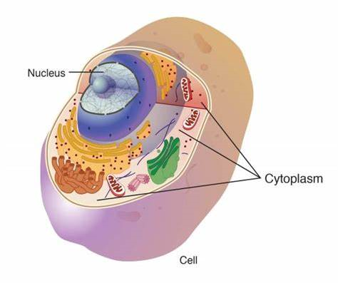
CYTOPLASM AND CELL ORGANELLES


INTRODUCTION:  Cytoplasm is a watery solution of minerals, gases, organic molecules, and cell organelles that is found between the cell membrane and the nucleus. Cytosol is the water portion of cytoplasm, and many chemical reactions take place within it. Cell organelles are intracellular structures, often bounded by their own membranes, that have specific functions in cellular metabolism.

ENDOPLASMIC RETICULUM:  The endoplasmic reticulum (ER) is an extensive network of membranous tubules that extend from the nuclear membrane to the cell membrane. Rough ER has numerous ribosomes on its surface, whereas smooth ER has no ribosomes. As a network of interconnected tunnels, the ER is a passage way for the transport of the materials necessary for cell function within the cell. These include proteins synthesized by the ribosomes on the rough ER, and lipids synthesized by the smooth ER. 

RIBOSOMES:  Ribosomes are very small structures made of protein and ribosomal RNA. Some are found on the surface of rough ER, while others float freely within the cytoplasm. Ribosomes are the site of protein synthesis. The proteins produced may be structural proteins such as collagen in the skin, enzymes, or hormones such as insulin that regulate cellular processes. These proteins may function within the cell or be secreted from the cell to be used elsewhere in the body. Our protein molecules are subject to damage, and some cellular proteins, especially regulatory proteins, may be needed just for a very short time. All such proteins must be destroyed, and this is the function of proteasomes. 

PROTEOSOMES:  A proteasome is a barrel-shaped organelle made of enzymes that cut protein molecules apart (protease enzymes). Proteins that are to be destroyed, that is, those no longer needed or those that are damaged or misfolded, are tagged by a protein called ubiquitin (sort of a cellular mop or broom) and carried into a proteasome. The protein is snipped into peptides or amino acids, which may be used again for protein synthesis on ribosomes. Proteasomes are particularly important during cell division and during embryonic development, when great changes are taking place very rapidly as cells become specialized. Many of our cells have secretory functions, that is, they produce specific products to be used elsewhere in tissues. 

GOLGI APPARATUS:  Secretion is one task of the Golgi apparatus, a series of flat, membranous sacs, somewhat like a stack of saucers. Carbohydrates are synthesized within the Golgi apparatus, and are packaged, along with other materials, for secretion from the cell. Proteins from the ribosomes or lipids from the smooth endoplasmic reticulum may also be secreted in this way. To secrete a substance, small sacs of the Golgi membrane break off and fuse with the cell membrane, releasing the substance to the exterior of the cell. This is exocytosis, exo meaning “to go out” of the cell. 

MITOCHONDRIA:  Mitochondria are oval or spherical organelles bounded by a double membrane. The inner membrane has folds called cristae. Within the mitochondria, the aerobic (oxygen-requiring) reactions of cell respiration take place. Therefore, mitochondria are the site of ATP (and hence energy) production. Cells that require large amounts of ATP, such as muscle cells, have many mitochondria to meet their need for energy. Mitochondria contain their own genes in a single DNA molecule and duplicate themselves when a cell divides. An individual’s mitochondrial DNA (mDNA) is of maternal origin, that is, from the mitochondria that were present in the ovum, or egg cell, which was then fertilized by a sperm cell. The mitochondria of the sperm cell usually do not enter the ovum during fertilization, because they are not found in the head of the sperm with the chromosomes.

LYSOSOMES:  Lysosomes are single-membrane structures that contain digestive enzymes. When certain white blood cells engulf bacteria, the bacteria are digested and destroyed by these lysosomal enzymes. Worn-out cell parts and dead cells are also digested by these enzymes. This is a beneficial process, and is necessary before tissue repair can begin. But it does have a disadvantage in that lysosomal digestion contributes to inflammation in damaged tissues. An excess of inflammation can start a vicious cycle, actually a positive feedback mechanism, that results in extensive tissue damage. Many of our cells are capable of dividing, or reproducing, themselves. 

CENTRIOLES:  Centrioles are a pair of rodshaped structures perpendicular to one another, located just outside the nucleus. Their function is to organize the spindle fibers during cell division. The spindle fibers are contracting proteins that pull the two sets of chromosomes apart, toward the ends of the original cell as it divides into two new cells. Each new cell then has a full set of chromosomes. 

CILIA AND FLAGELLA:  Cilia and flagella are mobile thread-like projections through the cell membrane; each is anchored by a basal body just within the membrane. Cilia are usually shorter than flagella, and an individual cell has many of them on its free surface. The cilia of a cell beat in unison and sweep materials across the cell surface. Cells lining the fallopian tubes, for example, have cilia to sweep the egg cell toward the uterus. The only human cell with a flagellum is the sperm cell. The flagellum provides motility, or movement, for the sperm cell. 

MICROVILLI:  Microvilli are folds of the cell membrane on the free surface of a cell.


RELATED;

1. CELLULAR METABOLISM  

2.  STUCTURE OF A CELL MEMBRANE  

3.  ENZYMES

4.  PROTEINS

5.  BODY TISSUES

REFERENCES


MOST FREQUENTLY READ